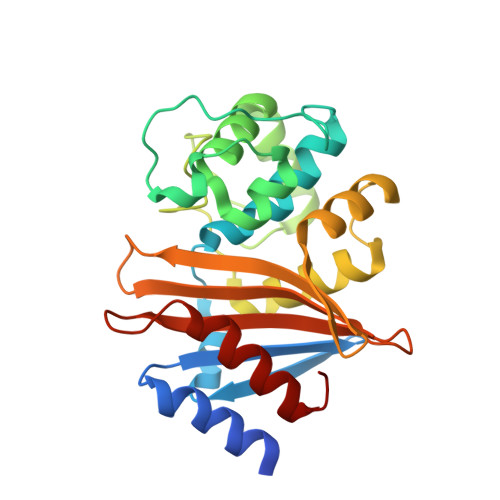
5L2F | pdb_00005l2f

High Resolution Structure of Acinetobacter baumannii beta-lactamase OXA-51 I129L/K83D bound to doripenem


Experimental Data Snapshot

  • Method: X-RAY DIFFRACTION
  • Resolution: 1.77 Å
  • R-Value Free: 
    0.192 (Depositor), 0.200 (DCC) 
  • R-Value Work: 
    0.175 (Depositor), 0.180 (DCC) 
  • R-Value Observed: 
    0.175 (Depositor) 

Starting Model: experimental
View more details

wwPDB Validation   3D Report Full Report


Ligand Structure Quality Assessment 

Created with Raphaël 2.3.0Worse 01 BetterLigand structure goodness of fit to experimental dataBest fitted 4J6Click on this verticalbar to view details

This is version 1.6 of the entry. See complete history


Literature

The structure of a doripenem-bound OXA-51 class D beta-lactamase variant with enhanced carbapenemase activity.

June, C.M.Muckenthaler, T.J.Schroder, E.C.Klamer, Z.L.Wawrzak, Z.Powers, R.A.Szarecka, A.Leonard, D.A.

(2016) Protein Sci 25: 2152-2163

  • DOI: https://doi.org/10.1002/pro.3040
  • Primary Citation of Related Structures:  
    5KZH, 5L2F

  • PubMed Abstract: 

    OXA-51 is a class D β-lactamase that is thought to be the native carbapenemase of Acinetobacter baumannii. Many variants of OXA-51 containing active site substitutions have been identified from A. baumannii isolates, and some of these substitutions increase hydrolytic activity toward carbapenem antibiotics. We have determined the high-resolution structures of apo OXA-51 and OXA-51 with one such substitution (I129L) with the carbapenem doripenem trapped in the active site as an acyl-intermediate. The structure shows that acyl-doripenem adopts an orientation very similar to carbapenem ligands observed in the active site of OXA-24/40 (doripenem) and OXA-23 (meropenem). In the OXA-51 variant/doripenem complex, the indole ring of W222 is oriented away from the doripenem binding site, thereby eliminating a clash that is predicted to occur in wildtype OXA-51. Similarly, in the OXA-51 variant complex, L129 adopts a different rotamer compared to I129 in wildtype OXA-51. This alternative position moves its side chain away from the hydroxyethyl moiety of doripenem and relieves another potential clash between the enzyme and carbapenem substrates. Molecular dynamics simulations of OXA-51 and OXA-51 I129L demonstrate that compared to isoleucine, a leucine at this position greatly favors a rotamer that accommodates the ligand. These results provide a molecular justification for how this substitution generates enhanced binding affinity for carbapenems, and therefore helps explain the prevalence of this substitution in clinical OXA-51 variants.


  • Organizational Affiliation

    Department of Chemistry, Grand Valley State University, Allendale, Michigan, 49401.


Macromolecules
Find similar proteins by:  (by identity cutoff)  |  3D Structure
Entity ID: 1
MoleculeChains Sequence LengthOrganismDetailsImage
Beta-lactamase
A, B, C, D
250Acinetobacter baumanniiMutation(s): 2 
Gene Names: oxa-51blaOXA-51AQ480_08190
UniProt
Find proteins for Q5QT35 (Acinetobacter baumannii)
Explore Q5QT35 
Go to UniProtKB:  Q5QT35
Entity Groups  
Sequence Clusters30% Identity50% Identity70% Identity90% Identity95% Identity100% Identity
UniProt GroupQ5QT35
Sequence Annotations
Expand
  • Reference Sequence
Small Molecules
Ligands 3 Unique
IDChains Name / Formula / InChI Key2D Diagram3D Interactions
4J6
Query on 4J6

Download Ideal Coordinates CCD File 
E [auth A],
H [auth B],
J [auth C],
N [auth D]
(4R,5S)-5-[(2S,3R)-3-hydroxy-1-oxobutan-2-yl]-4-methyl-3-({(3S,5S)-5-[(sulfamoylamino)methyl]pyrrolidin-3-yl}sulfanyl)-4,5-dihydro-1H-pyrrole-2-carboxylic acid
C15 H26 N4 O6 S2
QJKQOMNNBOTMCR-PQTSNVLCSA-N
GOL
Query on GOL

Download Ideal Coordinates CCD File 
G [auth A]
I [auth B]
L [auth C]
M [auth C]
P [auth D]
GLYCEROL
C3 H8 O3
PEDCQBHIVMGVHV-UHFFFAOYSA-N
ACT
Query on ACT

Download Ideal Coordinates CCD File 
F [auth A],
K [auth C],
O [auth D]
ACETATE ION
C2 H3 O2
QTBSBXVTEAMEQO-UHFFFAOYSA-M
Experimental Data & Validation

Experimental Data

  • Method: X-RAY DIFFRACTION
  • Resolution: 1.77 Å
  • R-Value Free:  0.192 (Depositor), 0.200 (DCC) 
  • R-Value Work:  0.175 (Depositor), 0.180 (DCC) 
  • R-Value Observed: 0.175 (Depositor) 
Space Group: P 21 21 21
Unit Cell:
Length ( Å )Angle ( ˚ )
a = 88.367α = 90
b = 92.149β = 90
c = 170.355γ = 90
Software Package:
Software NamePurpose
REFMACrefinement
HKL-2000data collection
HKL-2000data scaling
PHASERphasing

Structure Validation

View Full Validation Report


Ligand Structure Quality Assessment 

Created with Raphaël 2.3.0Worse 01 BetterLigand structure goodness of fit to experimental dataBest fitted 4J6Click on this verticalbar to view details

Entry History & Funding Information

Deposition Data


Funding OrganizationLocationGrant Number
National Institutes of Health/National Institute Of Allergy and Infectious Diseases (NIH/NIAID)United States1R15AI082416
National Institutes of Health/National Institute Of Allergy and Infectious Diseases (NIH/NIAID)United StatesR15AI094489

Revision History  (Full details and data files)

  • Version 1.0: 2016-09-28
    Type: Initial release
  • Version 1.1: 2016-10-05
    Changes: Database references
  • Version 1.2: 2016-12-07
    Changes: Database references
  • Version 1.3: 2017-09-27
    Changes: Author supporting evidence, Database references, Refinement description
  • Version 1.4: 2019-12-11
    Changes: Author supporting evidence
  • Version 1.5: 2023-10-04
    Changes: Data collection, Database references, Refinement description
  • Version 1.6: 2024-11-13
    Changes: Structure summary