5L3K | pdb_00005l3k

Structure of Mycobacterium thermoresistibile trehalose-6-phosphate synthase in a ternary complex with ADP and fructose-6-phosphate


Experimental Data Snapshot

  • Method: X-RAY DIFFRACTION
  • Resolution: 2.31 Å
  • R-Value Free: 
    0.213 (Depositor), 0.208 (DCC) 
  • R-Value Work: 
    0.153 (Depositor), 0.150 (DCC) 
  • R-Value Observed: 
    0.157 (Depositor) 

Starting Model: experimental
View more details

wwPDB Validation 3D Report Full Report

Validation slider image for 5L3K

Ligand Structure Quality Assessment 


This is version 1.4 of the entry. See complete history

Literature

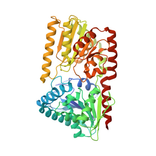
Mycobacterial OtsA Structures Unveil Substrate Preference Mechanism and Allosteric Regulation by 2-Oxoglutarate and 2-Phosphoglycerate.

Mendes, V.Acebron-Garcia-de-Eulate, M.Verma, N.Blaszczyk, M.Dias, M.V.B.Blundell, T.L.

(2019) mBio 10

  • DOI: https://doi.org/10.1128/mBio.02272-19
  • Primary Citation Related Structures: 
    5JIJ, 5JIO, 5K41, 5K42, 5K44, 5K5C, 5L3K

  • PubMed Abstract: 

    Trehalose is an essential disaccharide for mycobacteria and a key constituent of several cell wall glycolipids with fundamental roles in pathogenesis. Mycobacteria possess two pathways for trehalose biosynthesis. However, only the OtsAB pathway was found to be essential in Mycobacterium tuberculosis , with marked growth and virulence defects of OtsA mutants and strict essentiality of OtsB2. Here, we report the first mycobacterial OtsA structures from Mycobacterium thermoresistibile in both apo and ligand-bound forms. Structural information reveals three key residues in the mechanism of substrate preference that were further confirmed by site-directed mutagenesis. Additionally, we identify 2-oxoglutarate and 2-phosphoglycerate as allosteric regulators of OtsA. The structural analysis in this work strongly contributed to define the mechanisms for feedback inhibition, show different conformational states of the enzyme, and map a new allosteric site. IMPORTANCE Mycobacterial infections are a significant source of mortality worldwide, causing millions of deaths annually. Trehalose is a multipurpose disaccharide that plays a fundamental structural role in these organisms as a component of mycolic acids, a molecular hallmark of the cell envelope of mycobacteria. Here, we describe the first mycobacterial OtsA structures. We show mechanisms of substrate preference and show that OtsA is regulated allosterically by 2-oxoglutarate and 2-phosphoglycerate at an interfacial site. These results identify a new allosteric site and provide insight on the regulation of trehalose synthesis through the OtsAB pathway in mycobacteria.


  • Organizational Affiliation
    • Department of Biochemistry, University of Cambridge, Cambridge, United Kingdom vgm23@cam.ac.uk tom@cryst.bioc.cam.ac.uk.

Macromolecule Content 

  • Total Structure Weight: 442.92 kDa 
  • Atom Count: 29,978 
  • Modeled Residue Count: 3,597 
  • Deposited Residue Count: 3,896 
  • Unique protein chains: 1

Macromolecules

Find similar proteins by:|  3D Structure
Entity ID: 1
MoleculeChains  Sequence LengthOrganismDetailsImage
Alpha,alpha-trehalose-phosphate synthase
A, B, C, D, E
A, B, C, D, E, F, G, H
487Mycolicibacterium thermoresistibileMutation(s): 0 
Gene Names: RMCT_1906
EC: 2.4.1.347
UniProt
Find proteins for A0A117IMA6 (Mycolicibacterium thermoresistibile)
Explore A0A117IMA6 
Go to UniProtKB:  A0A117IMA6
Entity Groups
Sequence Clusters30% Identity50% Identity70% Identity90% Identity95% Identity100% Identity
UniProt GroupA0A117IMA6
Sequence Annotations
Expand
Reference Sequence

Small Molecules

Ligands 4 Unique
IDChains Name / Formula / InChI Key2D Diagram3D Interactions
ADP

Query on ADP


Download:Ideal Coordinates CCD File
AA [auth H]
I [auth A]
K [auth B]
N [auth C]
Q [auth D]
AA [auth H],
I [auth A],
K [auth B],
N [auth C],
Q [auth D],
U [auth E],
W [auth F],
Z [auth G]
ADENOSINE-5'-DIPHOSPHATE
C10 H15 N5 O10 P2
XTWYTFMLZFPYCI-KQYNXXCUSA-N
F6P

Query on F6P


Download:Ideal Coordinates CCD File
L [auth B],
O [auth C],
R [auth D],
V [auth E],
X [auth F]
6-O-phosphono-beta-D-fructofuranose
C6 H13 O9 P
BGWGXPAPYGQALX-ARQDHWQXSA-N
PO4

Query on PO4


Download:Ideal Coordinates CCD File
CA [auth H]
DA [auth H]
M [auth B]
S [auth D]
T [auth D]
CA [auth H],
DA [auth H],
M [auth B],
S [auth D],
T [auth D],
Y [auth F]
PHOSPHATE ION
O4 P
NBIIXXVUZAFLBC-UHFFFAOYSA-K
EDO

Query on EDO


Download:Ideal Coordinates CCD File
BA [auth H],
J [auth A],
P [auth C]
1,2-ETHANEDIOL
C2 H6 O2
LYCAIKOWRPUZTN-UHFFFAOYSA-N

Experimental Data & Validation

Experimental Data

  • Method: X-RAY DIFFRACTION
  • Resolution: 2.31 Å
  • R-Value Free:  0.213 (Depositor), 0.208 (DCC) 
  • R-Value Work:  0.153 (Depositor), 0.150 (DCC) 
  • R-Value Observed: 0.157 (Depositor) 
Space Group: P 62
Unit Cell:
Length ( Å )Angle ( ˚ )
a = 216.96α = 90
b = 216.96β = 90
c = 159.835γ = 120
Software Package:
Software NamePurpose
PHENIXrefinement
Aimlessdata scaling
PHASERphasing
PDB_EXTRACTdata extraction
XDSdata reduction

Structure Validation

View Full Validation Report


Ligand Structure Quality Assessment 


Entry History 

& Funding Information

Deposition Data


Funding OrganizationLocationGrant Number
Bill & Melinda Gates FoundationUnited States--

Revision History  (Full details and data files)

  • Version 1.0: 2017-06-07
    Type: Initial release
  • Version 1.1: 2017-09-13
    Changes: Author supporting evidence
  • Version 1.2: 2020-01-22
    Changes: Database references
  • Version 1.3: 2020-07-29
    Type: Remediation
    Reason: Carbohydrate remediation
    Changes: Data collection, Derived calculations, Structure summary
  • Version 1.4: 2024-01-10
    Changes: Data collection, Database references, Refinement description, Structure summary