5L70 | pdb_00005l70

CRYSTAL STRUCTURE OF HUMAN CARBONIC ANHYDRASE II IN COMPLEX WITH A QUINOLINE OLIGOAMIDE FOLDAMER


Experimental Data Snapshot

  • Method: X-RAY DIFFRACTION
  • Resolution: 2.20 Å
  • R-Value Free: 
    0.238 (Depositor), 0.236 (DCC) 
  • R-Value Work: 
    0.174 (Depositor), 0.181 (DCC) 
  • R-Value Observed: 
    0.177 (Depositor) 

Starting Model: experimental
View more details

wwPDB Validation 3D Report Full Report

Validation slider image for 5L70

This is version 4.2 of the entry. See complete history

Literature

CRYSTAL STRUCTURE OF HUMAN CARBONIC ANHYDRASE II IN COMPLEX WITH A QUINOLINE OLIGOAMIDE FOLDAMER

Vallade, M.Fischer, L.Langlois d'Estaintot, B.Granier, T.Huc, I.

To be published.

Macromolecule Content 

  • Total Structure Weight: 62.54 kDa 
  • Atom Count: 4,593 
  • Modeled Residue Count: 528 
  • Deposited Residue Count: 532 
  • Unique protein chains: 2

Macromolecules

Find similar proteins by:|  3D Structure
Entity ID: 1
MoleculeChains  Sequence LengthOrganismDetailsImage
Carbonic anhydrase 2
A, B
260Homo sapiensMutation(s): 0 
Gene Names: CA2
EC: 4.2.1.1 (PDB Primary Data), 4.2.1.69 (UniProt)
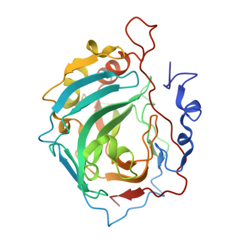
UniProt & NIH Common Fund Data Resources
Find proteins for P00918 (Homo sapiens)
Explore P00918 
Go to UniProtKB:  P00918
PHAROS:  P00918
GTEx:  ENSG00000104267 
Entity Groups
Sequence Clusters30% Identity50% Identity70% Identity90% Identity95% Identity100% Identity
UniProt GroupP00918
Sequence Annotations
Expand
Reference Sequence
Find similar proteins by:  Sequence   |   3D Structure  
Entity ID: 2
MoleculeChains  Sequence LengthOrganismDetailsImage
Aromatic foldamer
C, D
6synthetic constructMutation(s): 0 

Small Molecules

Ligands 2 Unique
IDChains Name / Formula / InChI Key2D Diagram3D Interactions
GOL

Query on GOL


Download:Ideal Coordinates CCD File
G [auth A],
O [auth A],
R [auth B],
U [auth B]
GLYCEROL
C3 H8 O3
PEDCQBHIVMGVHV-UHFFFAOYSA-N
ZN

Query on ZN


Download:Ideal Coordinates CCD File
E [auth A]
F [auth A]
H [auth A]
I [auth A]
J [auth A]
E [auth A],
F [auth A],
H [auth A],
I [auth A],
J [auth A],
K [auth A],
L [auth A],
M [auth A],
N [auth A],
P [auth B],
Q [auth B],
S [auth B],
T [auth B]
ZINC ION
Zn
PTFCDOFLOPIGGS-UHFFFAOYSA-N
Binding Affinity Annotations 
IDSourceBinding Affinity
4SO BindingDB:  5L70 Ki: min: 33, max: 260 (nM) from 6 assay(s)
Kd: 890 (nM) from 1 assay(s)
IC50: min: 880, max: 1.50e+4 (nM) from 2 assay(s)

Experimental Data & Validation

Experimental Data

  • Method: X-RAY DIFFRACTION
  • Resolution: 2.20 Å
  • R-Value Free:  0.238 (Depositor), 0.236 (DCC) 
  • R-Value Work:  0.174 (Depositor), 0.181 (DCC) 
  • R-Value Observed: 0.177 (Depositor) 
Space Group: P 1 21 1
Unit Cell:
Length ( Å )Angle ( ˚ )
a = 44.21α = 90
b = 83.37β = 99.98
c = 79.41γ = 90
Software Package:
Software NamePurpose
MxCuBEdata collection
SCALAdata scaling
REFMACrefinement
PDB_EXTRACTdata extraction
MOSFLMdata reduction
PHASERphasing

Structure Validation

View Full Validation Report


Entry History 

Deposition Data

Revision History  (Full details and data files)

  • Version 1.0: 2017-06-21
    Type: Initial release
  • Version 1.1: 2019-06-12
    Changes: Data collection, Database references, Structure summary
  • Version 2.0: 2023-11-15
    Changes: Advisory, Atomic model, Data collection, Database references, Derived calculations, Refinement description, Structure summary
  • Version 2.1: 2024-01-10
    Changes: Refinement description
  • Version 3.0: 2024-09-04
    Changes: Advisory, Atomic model, Data collection, Database references, Derived calculations, Non-polymer description, Other, Polymer sequence, Source and taxonomy, Structure summary
  • Version 4.0: 2024-10-02
    Changes: Atomic model, Data collection, Derived calculations, Non-polymer description, Structure summary
  • Version 4.1: 2024-11-20
    Changes: Structure summary
  • Version 4.2: 2025-06-11
    Changes: Data collection, Derived calculations