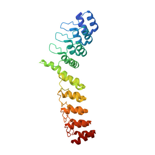
RCSB PDB - 5LE6: Crystal structure of DARPin-DARPin rigid fusion, variant DD_D12_09_D12

 5LE6

Crystal structure of DARPin-DARPin rigid fusion, variant DD_D12_09_D12


Experimental Data Snapshot

  • Method: X-RAY DIFFRACTION
  • Resolution: 1.80 Å
  • R-Value Free: 0.226 
  • R-Value Work: 0.192 
  • R-Value Observed: 0.194 

Starting Model: experimental
View more details

wwPDB Validation   3D Report Full Report


This is version 1.2 of the entry. See complete history


Literature

Rigidly connected multispecific artificial binders with adjustable geometries.

Wu, Y.Batyuk, A.Honegger, A.Brandl, F.Mittl, P.R.E.Pluckthun, A.

(2017) Sci Rep 7: 11217-11217

  • DOI: https://doi.org/10.1038/s41598-017-11472-x
  • Primary Citation of Related Structures:  
    5LE2, 5LE3, 5LE4, 5LE6, 5LE7, 5LE8, 5LE9, 5LEA, 5LEB, 5LEC, 5LED, 5LEE, 5LEL, 5LEM, 5LW2

  • PubMed Abstract: 

    Multivalent binding proteins can gain biological activities beyond what is inherent in the individual binders, by bringing together different target molecules, restricting their conformational flexibility or changing their subcellular localization. In this study, we demonstrate a method to build up rigid multivalent and multispecific scaffolds by exploiting the modular nature of a repeat protein scaffold and avoiding flexible linkers. We use DARPins (Designed Ankyrin Repeat Proteins), synthetic binding proteins based on the Ankyrin-repeat protein scaffold, as binding units. Their ease of in vitro selection, high production yield and stability make them ideal specificity-conferring building blocks for the design of more complex constructs. C- and N-terminal DARPin capping repeats were re-designed to be joined by a shared helix in such a way that rigid connector modules are formed. This allows us to join two or more DARPins in predefined geometries without compromising their binding affinities and specificities. Nine connector modules with distinct geometries were designed; for eight of these we were able to confirm the structure by X-ray crystallography, while only one did not crystallize. The bispecific constructs were all able to bind both target proteins simultaneously.


  • Organizational Affiliation

    Department of Biochemistry, University of Zürich, Winterthurerstrasse 190, CH-8057, Zürich, Switzerland.


Macromolecules
Find similar proteins by:  (by identity cutoff)  |  3D Structure
Entity ID: 1
MoleculeChains Sequence LengthOrganismDetailsImage
DD_D12_09_D12
A, B, C, D, E
324synthetic constructMutation(s): 0 
Entity Groups  
Sequence Clusters30% Identity50% Identity70% Identity90% Identity95% Identity100% Identity
Sequence Annotations
Expand
  • Reference Sequence
Small Molecules
Ligands 2 Unique
IDChains Name / Formula / InChI Key2D Diagram3D Interactions
SO4
Query on SO4

Download Ideal Coordinates CCD File 
L [auth D],
O [auth E],
Q [auth F]
SULFATE ION
O4 S
QAOWNCQODCNURD-UHFFFAOYSA-L
GOL
Query on GOL

Download Ideal Coordinates CCD File 
G [auth A]
H [auth A]
I [auth B]
J [auth B]
K [auth C]
GLYCEROL
C3 H8 O3
PEDCQBHIVMGVHV-UHFFFAOYSA-N
Experimental Data & Validation

Experimental Data

  • Method: X-RAY DIFFRACTION
  • Resolution: 1.80 Å
  • R-Value Free: 0.226 
  • R-Value Work: 0.192 
  • R-Value Observed: 0.194 
  • Space Group: P 61 2 2
Unit Cell:
Length ( Å )Angle ( ˚ )
a = 145.12α = 90
b = 145.12β = 90
c = 376.76γ = 120
Software Package:
Software NamePurpose
XDSdata reduction
XSCALEdata scaling
PHASERphasing
PHENIXrefinement

Structure Validation

View Full Validation Report


Entry History & Funding Information

Deposition Data


Funding OrganizationLocationGrant Number
Swiss National Science FoundationSwitzerland310030B_166676
European Research CouncilNEXTBINDERS

Revision History  (Full details and data files)

  • Version 1.0: 2017-08-02
    Type: Initial release
  • Version 1.1: 2019-03-20
    Changes: Data collection, Database references
  • Version 1.2: 2024-01-10
    Changes: Data collection, Database references, Refinement description