First-in-Class Chemical Probes against Poly(ADP-ribose) Glycohydrolase (PARG) Inhibit DNA Repair with Differential Pharmacology to Olaparib.
James, D.I., Smith, K.M., Jordan, A.M., Fairweather, E.E., Griffiths, L.A., Hamilton, N.S., Hitchin, J.R., Hutton, C.P., Jones, S., Kelly, P., McGonagle, A.E., Small, H., Stowell, A.I., Tucker, J., Waddell, I.D., Waszkowycz, B., Ogilvie, D.J.(2016) ACS Chem Biol 11: 3179-3190
- PubMed: 27689388 Search on PubMed
- DOI: https://doi.org/10.1021/acschembio.6b00609
- Primary Citation Related Structures:
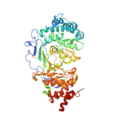
5LHB - PubMed Abstract:
The enzyme poly(ADP-ribose) glycohydrolase (PARG) performs a critical role in the repair of DNA single strand breaks (SSBs). However, a detailed understanding of its mechanism of action has been hampered by a lack of credible, cell-active chemical probes. Herein, we demonstrate inhibition of PARG with a small molecule, leading to poly(ADP-ribose) (PAR) chain persistence in intact cells. Moreover, we describe two advanced, and chemically distinct, cell-active tool compounds with convincing on-target pharmacology and selectivity. Using one of these tool compounds, we demonstrate pharmacology consistent with PARG inhibition. Further, while the roles of PARG and poly(ADP-ribose) polymerase (PARP) are closely intertwined, we demonstrate that the pharmacology of a PARG inhibitor differs from that observed with the more thoroughly studied PARP inhibitor olaparib. We believe that these tools will facilitate a wider understanding of this important component of DNA repair and may enable the development of novel therapeutic agents exploiting the critical dependence of tumors on the DNA damage response (DDR).
- Drug Discovery Unit, Cancer Research UK Manchester Institute, University of Manchester , Wilmslow Road, Manchester, M20 4BX, United Kingdom.
Organizational Affiliation: