5LQY | pdb_00005lqy

Structure of F-ATPase from Pichia angusta, in state2


Experimental Data Snapshot

  • Method: ELECTRON MICROSCOPY
  • Resolution: 7.80 Å
  • Aggregation State: PARTICLE 
  • Reconstruction Method: SINGLE PARTICLE 

wwPDB Validation 3D Report Full Report

Validation slider image for 5LQY

This is version 1.5 of the entry. See complete history. 

Literature

Structure of the mitochondrial ATP synthase fromPichia angustadetermined by electron cryo-microscopy.

Vinothkumar, K.R., Montgomery, M.G., Liu, S., Walker, J.E.

(2016) Proc Natl Acad Sci U S A 113: 12709-12714

  • DOI: https://doi.org/10.1073/pnas.1615902113
  • Primary Citation Related Structures: 
    5LQX, 5LQY, 5LQZ

  • PubMed Abstract: 

    The structure of the intact monomeric ATP synthase from the fungus, Pichia angusta , has been solved by electron cryo-microscopy. The structure provides insights into the mechanical coupling of the transmembrane proton motive force across mitochondrial membranes in the synthesis of ATP. This mechanism requires a strong and integral stator, consisting of the catalytic α 3 β 3 -domain, peripheral stalk, and, in the membrane domain, subunit a and associated supernumerary subunits, kept in contact with the rotor turning at speeds up to 350 Hz. The stator's integrity is ensured by robust attachment of both the oligomycin sensitivity conferral protein (OSCP) to the catalytic domain and the membrane domain of subunit b to subunit a. The ATP8 subunit provides an additional brace between the peripheral stalk and subunit a. At the junction between the OSCP and the apparently stiff, elongated α-helical b-subunit and associated d- and h-subunits, an elbow or joint allows the stator to bend to accommodate lateral movements during the activity of the catalytic domain. The stator may also apply lateral force to help keep the static a-subunit and rotating c 10 -ring together. The interface between the c 10 -ring and the a-subunit contains the transmembrane pathway for protons, and their passage across the membrane generates the turning of the rotor. The pathway has two half-channels containing conserved polar residues provided by a bundle of four α-helices inclined at ∼30° to the plane of the membrane, similar to those described in other species. The structure provides more insights into the workings of this amazing machine.


  • Organizational Affiliation: 
    • The Medical Research Council Laboratory of Molecular Biology, Cambridge Biomedical Campus, Cambridge CB2 0QH, United Kingdom.

Macromolecule Content 

  • Total Structure Weight: 557.71 kDa 
  • Atom Count: 24,170 
  • Modeled Residue Count: 4,881 
  • Deposited Residue Count: 5,223 
  • Unique protein chains: 17

Macromolecules

Find similar proteins by:|  3D Structure
Entity ID: 1
MoleculeChains  Sequence LengthOrganismDetailsImage
ATP synthase subunit fA [auth 1]30Ogataea angustaMutation(s): 0 
Sequence Annotations
Expand
Reference Sequence
Find similar proteins by:  Sequence   |   3D Structure  
Entity ID: 2
MoleculeChains  Sequence LengthOrganismDetailsImage
ATP synthase subunit AAP1B [auth 2]25Ogataea angustaMutation(s): 0 
Sequence Annotations
Expand
Reference Sequence
Find similar proteins by:  Sequence   |   3D Structure  
Entity ID: 3
MoleculeChains  Sequence LengthOrganismDetailsImage
ATP synthase subunit aC [auth 3]17Ogataea angustaMutation(s): 0 
Sequence Annotations
Expand
Reference Sequence
Find similar proteins by:|  3D Structure
Entity ID: 4
MoleculeChains  Sequence LengthOrganismDetailsImage
ATP synthase subunit bD [auth 4]27Ogataea angustaMutation(s): 0 
Sequence Annotations
Expand
Reference Sequence
Find similar proteins by:|  3D Structure
Entity ID: 5
MoleculeChains  Sequence LengthOrganismDetailsImage
ATP synthase alpha subunitE [auth A],
F [auth B],
G [auth C]
510Ogataea angustaMutation(s): 0 
Entity Groups
Sequence Clusters30% Identity50% Identity70% Identity90% Identity95% Identity100% Identity
Sequence Annotations
Expand
Reference Sequence
Find similar proteins by:|  3D Structure
Entity ID: 6
MoleculeChains  Sequence LengthOrganismDetailsImage
ATP synthase beta subunitH [auth D],
I [auth E],
J [auth F]
476Ogataea angustaMutation(s): 0 
Entity Groups
Sequence Clusters30% Identity50% Identity70% Identity90% Identity95% Identity100% Identity
Sequence Annotations
Expand
Reference Sequence
Find similar proteins by:|  3D Structure
Entity ID: 7
MoleculeChains  Sequence LengthOrganismDetailsImage
ATP synthase gamma subunitK [auth G]269Ogataea angustaMutation(s): 0 
Entity Groups
Sequence Clusters30% Identity50% Identity70% Identity90% Identity95% Identity100% Identity
Sequence Annotations
Expand
Reference Sequence
Find similar proteins by:|  3D Structure
Entity ID: 8
MoleculeChains  Sequence LengthOrganismDetailsImage
ATP synthase delta subunitL [auth H]138Ogataea angustaMutation(s): 0 
Entity Groups
Sequence Clusters30% Identity50% Identity70% Identity90% Identity95% Identity100% Identity
Sequence Annotations
Expand
Reference Sequence
Find similar proteins by:|  3D Structure
Entity ID: 9
MoleculeChains  Sequence LengthOrganismDetailsImage
ATP synthase epsilon subunitM [auth I]63Ogataea angustaMutation(s): 0 
Entity Groups
Sequence Clusters30% Identity50% Identity70% Identity90% Identity95% Identity100% Identity
Sequence Annotations
Expand
Reference Sequence
Find similar proteins by:|  3D Structure
Entity ID: 10
MoleculeChains  Sequence LengthOrganismDetailsImage
ATP synthase inhibitor protein IF1N [auth J]66Bos taurusMutation(s): 0 
Entity Groups
Sequence Clusters30% Identity50% Identity70% Identity90% Identity95% Identity100% Identity
Sequence Annotations
Expand
Reference Sequence
Find similar proteins by:|  3D Structure
Entity ID: 11
MoleculeChains  Sequence LengthOrganismDetailsImage
ATP synthase c subunit76Ogataea angustaMutation(s): 0 
Entity Groups
Sequence Clusters30% Identity50% Identity70% Identity90% Identity95% Identity100% Identity
Sequence Annotations
Expand
Reference Sequence
Find similar proteins by:|  3D Structure
Entity ID: 12
MoleculeChains  Sequence LengthOrganismDetailsImage
ATP synthase OSCP subunitY [auth U]194Ogataea angustaMutation(s): 0 
Entity Groups
Sequence Clusters30% Identity50% Identity70% Identity90% Identity95% Identity100% Identity
Sequence Annotations
Expand
Reference Sequence
Find similar proteins by:|  3D Structure
Entity ID: 13
MoleculeChains  Sequence LengthOrganismDetailsImage
ATP synthase subunit bZ [auth V]204Ogataea angustaMutation(s): 0 
Entity Groups
Sequence Clusters30% Identity50% Identity70% Identity90% Identity95% Identity100% Identity
Sequence Annotations
Expand
Reference Sequence
Find similar proteins by:|  3D Structure
Entity ID: 14
MoleculeChains  Sequence LengthOrganismDetailsImage
ATP synthase subunit dAA [auth W]155Ogataea angustaMutation(s): 0 
Entity Groups
Sequence Clusters30% Identity50% Identity70% Identity90% Identity95% Identity100% Identity
Sequence Annotations
Expand
Reference Sequence
Find similar proteins by:  Sequence   |   3D Structure  
Entity ID: 15
MoleculeChains  Sequence LengthOrganismDetailsImage
ATP synthase subunit hBA [auth X]21Ogataea angustaMutation(s): 0 
Sequence Annotations
Expand
Reference Sequence
Find similar proteins by:|  3D Structure
Entity ID: 16
MoleculeChains  Sequence LengthOrganismDetailsImage
ATP synthase subunit aCA [auth Y]252Ogataea angustaMutation(s): 0 
Entity Groups
Sequence Clusters30% Identity50% Identity70% Identity90% Identity95% Identity100% Identity
Sequence Annotations
Expand
Reference Sequence
Find similar proteins by:|  3D Structure
Entity ID: 17
MoleculeChains  Sequence LengthOrganismDetailsImage
ATP synthase subunit aDA [auth Z]44Ogataea angustaMutation(s): 0 
Sequence Annotations
Expand
Reference Sequence

Small Molecules

Ligands 3 Unique
IDChains Name / Formula / InChI Key2D Diagram3D Interactions
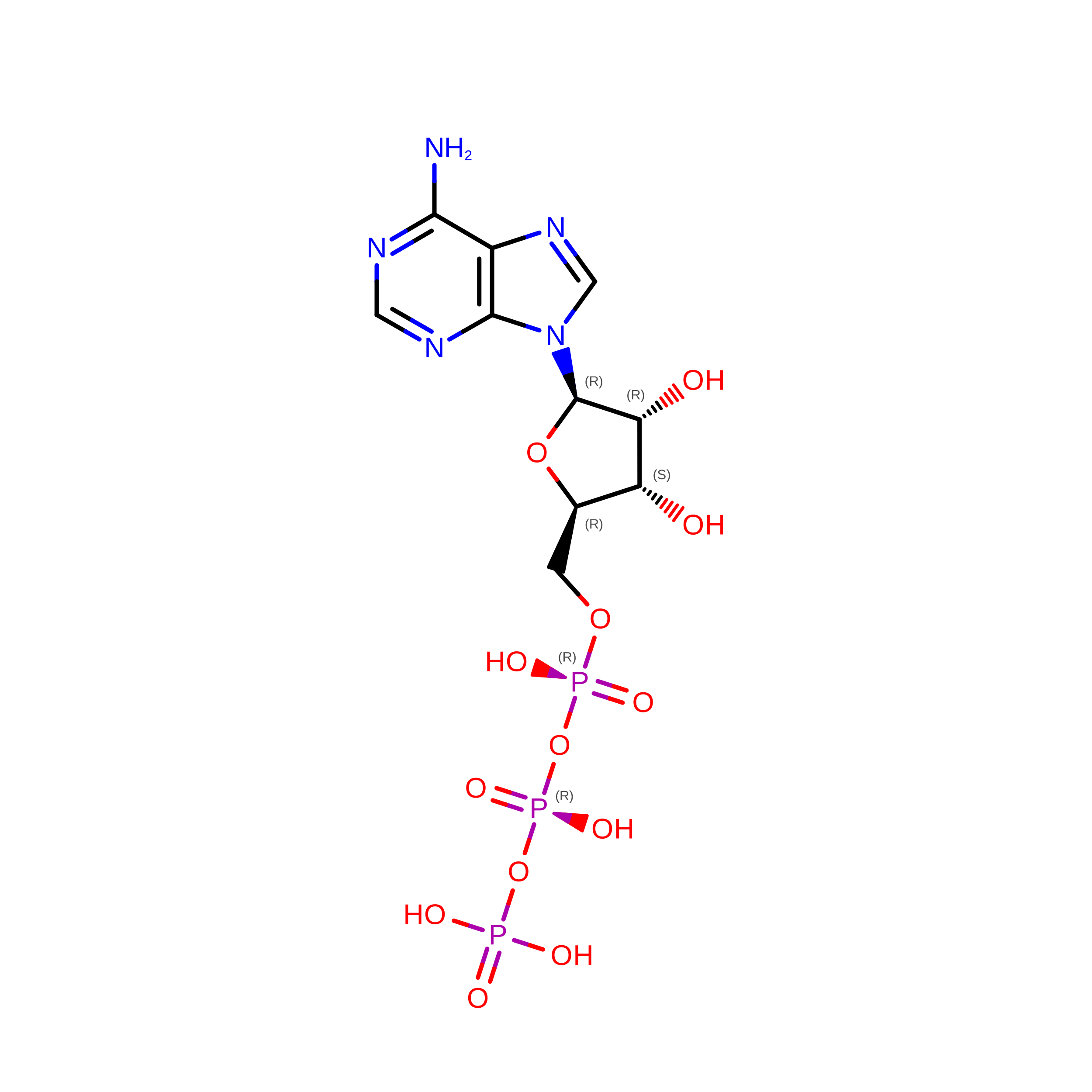
ATP

Query on ATP


Download:Ideal Coordinates CCD File
EA [auth A]ADENOSINE-5'-TRIPHOSPHATE
C10 H16 N5 O13 P3
ZKHQWZAMYRWXGA-KQYNXXCUSA-N
ADP

Query on ADP


Download:Ideal Coordinates CCD File
GA [auth B],
IA [auth C],
KA [auth D],
MA [auth E],
NA [auth F]
ADENOSINE-5'-DIPHOSPHATE
C10 H15 N5 O10 P2
XTWYTFMLZFPYCI-KQYNXXCUSA-N
MG

Query on MG


Download:Ideal Coordinates CCD File
FA [auth A],
HA [auth B],
JA [auth C],
LA [auth D],
OA [auth F]
MAGNESIUM ION
Mg
JLVVSXFLKOJNIY-UHFFFAOYSA-N
Non-standard Monomers  1 Unique
IDChains TypeFormula2D Diagram
UNK
Query on UNK
L-PEPTIDE LINKINGC4 H9 N O2

Experimental Data & Validation

Experimental Data

  • Method: ELECTRON MICROSCOPY
  • Resolution: 7.80 Å
  • Aggregation State: PARTICLE 
  • Reconstruction Method: SINGLE PARTICLE 
EM Software:
TaskSoftware PackageVersion
RECONSTRUCTIONRELION1.4

Structure Validation

View Full Validation Report


Entry History 

Deposition Data

Revision History  (Full details and data files)

  • Version 1.0: 2016-11-16
    Type: Initial release
  • Version 1.1: 2017-08-02
    Changes: Data collection, Experimental preparation
  • Version 1.2: 2018-01-24
    Changes: Source and taxonomy
  • Version 1.3: 2018-01-31
    Changes: Structure summary
  • Version 1.4: 2018-09-12
    Changes: Data collection, Database references
  • Version 1.5: 2024-05-15
    Changes: Data collection, Database references