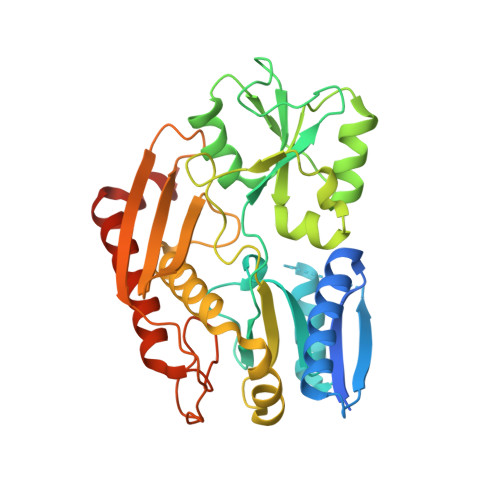
5M7F

Human porphobilinogen deaminase in complex with DPM cofactor


Experimental Data Snapshot

  • Method: X-RAY DIFFRACTION
  • Resolution: 2.78 Å
  • R-Value Free: 0.260 
  • R-Value Work: 0.220 
  • R-Value Observed: 0.222 

Starting Model: experimental
View more details

wwPDB Validation   3D Report Full Report


Ligand Structure Quality Assessment 


This is version 1.2 of the entry. See complete history


Literature

Structural basis of pyrrole polymerization in human porphobilinogen deaminase.

Pluta, P.Roversi, P.Bernardo-Seisdedos, G.Rojas, A.L.Cooper, J.B.Gu, S.Pickersgill, R.W.Millet, O.

(2018) Biochim Biophys Acta Gen Subj 1862: 1948-1955

  • DOI: https://doi.org/10.1016/j.bbagen.2018.06.013
  • Primary Citation of Related Structures:  
    5M6R, 5M7F

  • PubMed Abstract: 

    Human porphobilinogen deaminase (PBGD), the third enzyme in the heme pathway, catalyzes four times a single reaction to convert porphobilinogen into hydroxymethylbilane. Remarkably, PBGD employs a single active site during the process, with a distinct yet chemically equivalent bond formed each time. The four intermediate complexes of the enzyme have been biochemically validated and they can be isolated but they have never been structurally characterized other than the apo- and holo-enzyme bound to the cofactor. We present crystal structures for two human PBGD intermediates: PBGD loaded with the cofactor and with the reaction intermediate containing two additional substrate pyrrole rings. These results, combined with SAXS and NMR experiments, allow us to propose a mechanism for the reaction progression that requires less structural rearrangements than previously suggested: the enzyme slides a flexible loop over the growing-product active site cavity. The structures and the mechanism proposed for this essential reaction explain how a set of missense mutations result in acute intermittent porphyria.


  • Organizational Affiliation

    Protein Stability and Inherited Disease Laboratory, CIC bioGUNE, Derio, Bizkaia 48160, Spain.


Macromolecules
Find similar proteins by:  (by identity cutoff)  |  3D Structure
Entity ID: 1
MoleculeChains Sequence LengthOrganismDetailsImage
Porphobilinogen deaminase
A, B
365Homo sapiensMutation(s): 0 
Gene Names: HMBSPBGDUPS
EC: 2.5.1.61
UniProt & NIH Common Fund Data Resources
Find proteins for P08397 (Homo sapiens)
Explore P08397 
Go to UniProtKB:  P08397
PHAROS:  P08397
GTEx:  ENSG00000256269 
Entity Groups  
Sequence Clusters30% Identity50% Identity70% Identity90% Identity95% Identity100% Identity
UniProt GroupP08397
Sequence Annotations
Expand
  • Reference Sequence
Small Molecules
Ligands 2 Unique
IDChains Name / Formula / InChI Key2D Diagram3D Interactions
DPM
Query on DPM

Download Ideal Coordinates CCD File 
C [auth A],
G [auth B]
3-[5-{[3-(2-carboxyethyl)-4-(carboxymethyl)-5-methyl-1H-pyrrol-2-yl]methyl}-4-(carboxymethyl)-1H-pyrrol-3-yl]propanoic acid
C20 H24 N2 O8
LCAXMKQKEYTFDM-UHFFFAOYSA-N
SO4
Query on SO4

Download Ideal Coordinates CCD File 
D [auth A]
E [auth A]
F [auth A]
H [auth B]
I [auth B]
D [auth A],
E [auth A],
F [auth A],
H [auth B],
I [auth B],
J [auth B]
SULFATE ION
O4 S
QAOWNCQODCNURD-UHFFFAOYSA-L
Experimental Data & Validation

Experimental Data

  • Method: X-RAY DIFFRACTION
  • Resolution: 2.78 Å
  • R-Value Free: 0.260 
  • R-Value Work: 0.220 
  • R-Value Observed: 0.222 
  • Space Group: P 1 21 1
Unit Cell:
Length ( Å )Angle ( ˚ )
a = 70.42α = 90
b = 80.95β = 95.7
c = 76.22γ = 90
Software Package:
Software NamePurpose
BUSTERrefinement
XDSdata reduction
Aimlessdata scaling
MOLREPphasing

Structure Validation

View Full Validation Report


Ligand Structure Quality Assessment 


Entry History & Funding Information

Deposition Data


Funding OrganizationLocationGrant Number
Spain--

Revision History  (Full details and data files)

  • Version 1.0: 2017-11-15
    Type: Initial release
  • Version 1.1: 2019-05-29
    Changes: Data collection, Database references
  • Version 1.2: 2024-01-17
    Changes: Data collection, Database references, Refinement description