5MM0 | pdb_00005mm0

Dolichyl phosphate mannose synthase in complex with GDP-mannose and Mn2+ (donor complex)


Experimental Data Snapshot

  • Method: X-RAY DIFFRACTION
  • Resolution: 2.30 Å
  • R-Value Free: 
    0.258 (Depositor), 0.260 (DCC) 
  • R-Value Work: 
    0.219 (Depositor), 0.227 (DCC) 
  • R-Value Observed: 
    0.222 (Depositor) 

Starting Model: experimental
View more details

wwPDB Validation 3D Report Full Report

Validation slider image for 5MM0

Ligand Structure Quality Assessment 


This is version 1.1 of the entry. See complete history

Literature

Structural basis for dolichylphosphate mannose biosynthesis.

Gandini, R.Reichenbach, T.Tan, T.C.Divne, C.

(2017) Nat Commun 8: 120-120

  • DOI: https://doi.org/10.1038/s41467-017-00187-2
  • Primary Citation Related Structures: 
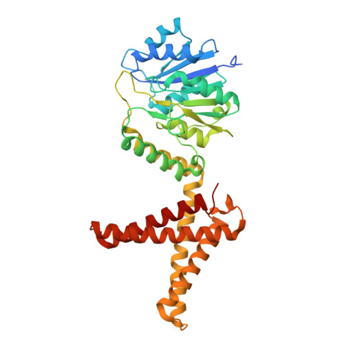
    5MLZ, 5MM0, 5MM1

  • PubMed Abstract: 

    Protein glycosylation is a critical protein modification. In biogenic membranes of eukaryotes and archaea, these reactions require activated mannose in the form of the lipid conjugate dolichylphosphate mannose (Dol-P-Man). The membrane protein dolichylphosphate mannose synthase (DPMS) catalyzes the reaction whereby mannose is transferred from GDP-mannose to the dolichol carrier Dol-P, to yield Dol-P-Man. Failure to produce or utilize Dol-P-Man compromises organism viability, and in humans, several mutations in the human dpm1 gene lead to congenital disorders of glycosylation (CDG). Here, we report three high-resolution crystal structures of archaeal DPMS from Pyrococcus furiosus, in complex with nucleotide, donor, and glycolipid product. The structures offer snapshots along the catalytic cycle, and reveal how lipid binding couples to movements of interface helices, metal binding, and acceptor loop dynamics to control critical events leading to Dol-P-Man synthesis. The structures also rationalize the loss of dolichylphosphate mannose synthase function in dpm1-associated CDG.The generation of glycolipid dolichylphosphate mannose (Dol-P-Man) is a critical step for protein glycosylation and GPI anchor synthesis. Here the authors report the structure of dolichylphosphate mannose synthase in complex with bound nucleotide and donor to provide insight into the mechanism of Dol-P-Man synthesis.


  • Organizational Affiliation
    • School of Biotechnology, KTH Royal Institute of Technology, S-10691, Stockholm, Sweden.

Macromolecule Content 

  • Total Structure Weight: 44.53 kDa 
  • Atom Count: 3,008 
  • Modeled Residue Count: 355 
  • Deposited Residue Count: 374 
  • Unique protein chains: 1

Macromolecules

Find similar proteins by:|  3D Structure
Entity ID: 1
MoleculeChains  Sequence LengthOrganismDetailsImage
Dolichol monophosphate mannose synthase374Pyrococcus furiosus DSM 3638Mutation(s): 0 
Gene Names: PF0058
EC: 2.4.1.83
Membrane Entity: Yes 
UniProt
Find proteins for Q8U4M3 (Pyrococcus furiosus (strain ATCC 43587 / DSM 3638 / JCM 8422 / Vc1))
Explore Q8U4M3 
Go to UniProtKB:  Q8U4M3
Entity Groups
Sequence Clusters30% Identity50% Identity70% Identity90% Identity95% Identity100% Identity
UniProt GroupQ8U4M3
Sequence Annotations
Expand
Reference Sequence

Small Molecules

Ligands 4 Unique
IDChains Name / Formula / InChI Key2D Diagram3D Interactions
GDD

Query on GDD


Download:Ideal Coordinates CCD File
B [auth A]GUANOSINE-5'-DIPHOSPHATE-ALPHA-D-MANNOSE
C16 H25 N5 O16 P2
MVMSCBBUIHUTGJ-GDJBGNAASA-N
LDA

Query on LDA


Download:Ideal Coordinates CCD File
E [auth A],
F [auth A],
G [auth A],
H [auth A],
I [auth A]
LAURYL DIMETHYLAMINE-N-OXIDE
C14 H31 N O
SYELZBGXAIXKHU-UHFFFAOYSA-N
MN

Query on MN


Download:Ideal Coordinates CCD File
C [auth A]MANGANESE (II) ION
Mn
WAEMQWOKJMHJLA-UHFFFAOYSA-N
CL

Query on CL


Download:Ideal Coordinates CCD File
D [auth A]CHLORIDE ION
Cl
VEXZGXHMUGYJMC-UHFFFAOYSA-M

Experimental Data & Validation

Experimental Data

  • Method: X-RAY DIFFRACTION
  • Resolution: 2.30 Å
  • R-Value Free:  0.258 (Depositor), 0.260 (DCC) 
  • R-Value Work:  0.219 (Depositor), 0.227 (DCC) 
  • R-Value Observed: 0.222 (Depositor) 
Space Group: C 2 2 21
Unit Cell:
Length ( Å )Angle ( ˚ )
a = 90.67α = 90
b = 146.32β = 90
c = 95.09γ = 90
Software Package:
Software NamePurpose
PHENIXrefinement
XDSdata reduction
XSCALEdata scaling
PHENIXphasing

Structure Validation

View Full Validation Report


Ligand Structure Quality Assessment 


Entry History 

& Funding Information

Deposition Data


Funding OrganizationLocationGrant Number
Swedish Research CouncilSweden2011-5768
Swedish Research CouncilSweden2013-5717
Swedish Research CouncilSweden2012-915

Revision History  (Full details and data files)

  • Version 1.0: 2017-08-09
    Type: Initial release
  • Version 1.1: 2024-01-17
    Changes: Data collection, Database references, Refinement description