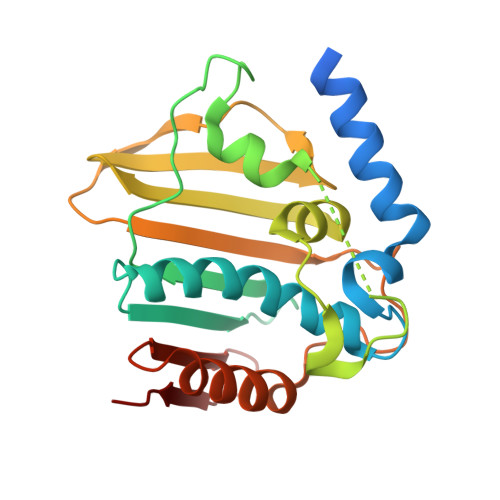
5MMN

E. coli DNA Gyrase B 24 kDa ATPase domain in complex with 1-ethyl-3-[8-methyl-5-(2-methyl-pyridin-4-yl)-isoquinolin-3-yl]-urea


Experimental Data Snapshot

  • Method: X-RAY DIFFRACTION
  • Resolution: 1.90 Å
  • R-Value Free: 0.227 
  • R-Value Work: 0.188 
  • R-Value Observed: 0.190 

wwPDB Validation   3D Report Full Report


Ligand Structure Quality Assessment 


This is version 1.2 of the entry. See complete history


Literature

Discovery and Optimization of Isoquinoline Ethyl Ureas as Antibacterial Agents.

Panchaud, P.Bruyere, T.Blumstein, A.C.Bur, D.Chambovey, A.Ertel, E.A.Gude, M.Hubschwerlen, C.Jacob, L.Kimmerlin, T.Pfeifer, T.Prade, L.Seiler, P.Ritz, D.Rueedi, G.

(2017) J Med Chem 60: 3755-3775

  • DOI: https://doi.org/10.1021/acs.jmedchem.6b01834
  • Primary Citation of Related Structures:  
    5MMN, 5MMO, 5MMP

  • PubMed Abstract: 

    Our strategy to combat resistant bacteria consisted of targeting the GyrB/ParE ATP-binding sites located on bacterial DNA gyrase and topoisomerase IV and not utilized by marketed antibiotics. Screening around the minimal ethyl urea binding motif led to the identification of isoquinoline ethyl urea 13 as a promising starting point for fragment evolution. The optimization was guided by structure-based design and focused on antibacterial activity in vitro and in vivo, culminating in the discovery of unprecedented substituents able to interact with conserved residues within the ATP-binding site. A detailed characterization of the lead compound highlighted the potential for treatment of the problematic fluoroquinolone-resistant MRSA, VRE, and S. pneumoniae, and the possibility to offer patients an intravenous-to-oral switch therapy was supported by the identification of a suitable prodrug concept. Eventually, hERG K-channel block was identified as the main limitation of this chemical series, and efforts toward its minimization are reported.


  • Organizational Affiliation

    Actelion Pharmaceuticals Ltd. , Gewerbestrasse 16, CH-4123 Allschwil, Switzerland.


Macromolecules
Find similar proteins by:  (by identity cutoff)  |  3D Structure
Entity ID: 1
MoleculeChains Sequence LengthOrganismDetailsImage
DNA gyrase subunit B234Escherichia coli O157:H7Mutation(s): 0 
Gene Names: gyrBZ5190ECs4634
EC: 5.99.1.3
UniProt
Find proteins for P0AES7 (Escherichia coli O157:H7)
Explore P0AES7 
Go to UniProtKB:  P0AES7
Entity Groups  
Sequence Clusters30% Identity50% Identity70% Identity90% Identity95% Identity100% Identity
UniProt GroupP0AES7
Sequence Annotations
Expand
  • Reference Sequence
Small Molecules
Ligands 1 Unique
IDChains Name / Formula / InChI Key2D Diagram3D Interactions
O54
Query on O54

Download Ideal Coordinates CCD File 
B [auth A]1-ethyl-3-[8-methyl-5-(2-methylpyridin-4-yl)isoquinolin-3-yl]urea
C19 H20 N4 O
ITGDBTWKMCUOIO-UHFFFAOYSA-N
Experimental Data & Validation

Experimental Data

  • Method: X-RAY DIFFRACTION
  • Resolution: 1.90 Å
  • R-Value Free: 0.227 
  • R-Value Work: 0.188 
  • R-Value Observed: 0.190 
  • Space Group: P 31 2 1
Unit Cell:
Length ( Å )Angle ( ˚ )
a = 71.52α = 90
b = 71.52β = 90
c = 78.95γ = 120
Software Package:
Software NamePurpose
REFMACrefinement
XDSdata reduction
XSCALEdata scaling
PHASERphasing

Structure Validation

View Full Validation Report


Ligand Structure Quality Assessment 


Entry History 

Revision History  (Full details and data files)

  • Version 1.0: 2017-04-26
    Type: Initial release
  • Version 1.1: 2017-05-24
    Changes: Database references
  • Version 1.2: 2024-05-08
    Changes: Data collection, Database references