5MTR | pdb_00005mtr

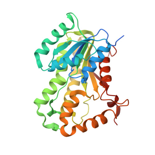
Crystal structure of M. tuberculosis InhA inhibited by PT512


Experimental Data Snapshot

  • Method: X-RAY DIFFRACTION
  • Resolution: 2.00 Å
  • R-Value Free: 
    0.234 (Depositor), 0.230 (DCC) 
  • R-Value Work: 
    0.205 (Depositor), 0.200 (DCC) 
  • R-Value Observed: 
    0.206 (Depositor) 

Starting Model: experimental
View more details

wwPDB Validation   3D Report Full Report


Ligand Structure Quality Assessment 

Created with Raphaël 2.3.0Worse 01 BetterLigand structure goodness of fit to experimental dataBest fitted NADClick on this verticalbar to view detailsBest fitted XT0Click on this verticalbar to view details

This is version 1.3 of the entry. See complete history


Literature

Evaluating the Contribution of Transition-State Destabilization to Changes in the Residence Time of Triazole-Based InhA Inhibitors.

Spagnuolo, L.A.Eltschkner, S.Yu, W.Daryaee, F.Davoodi, S.Knudson, S.E.Allen, E.K.Merino, J.Pschibul, A.Moree, B.Thivalapill, N.Truglio, J.J.Salafsky, J.Slayden, R.A.Kisker, C.Tonge, P.J.

(2017) J Am Chem Soc 139: 3417-3429

  • DOI: https://doi.org/10.1021/jacs.6b11148
  • Primary Citation of Related Structures:  
    5MTP, 5MTQ, 5MTR, 5UGS, 5UGT, 5UGU

  • PubMed Abstract: 

    A critical goal of lead compound selection and optimization is to maximize target engagement while minimizing off-target binding. Since target engagement is a function of both the thermodynamics and kinetics of drug-target interactions, it follows that the structures of both the ground states and transition states on the binding reaction coordinate are needed to rationally modulate the lifetime of the drug-target complex. Previously, we predicted the structure of the rate-limiting transition state that controlled the time-dependent inhibition of the enoyl-ACP reductase InhA. This led to the discovery of a triazole-containing diphenyl ether with an increased residence time on InhA due to transition-state destabilization rather than ground-state stabilization. In the present work, we evaluate the inhibition of InhA by 14 triazole-based diphenyl ethers and use a combination of enzyme kinetics and X-ray crystallography to generate a structure-kinetic relationship for time-dependent binding. We show that the triazole motif slows the rate of formation for the final drug-target complex by up to 3 orders of magnitude. In addition, we identify a novel inhibitor with a residence time on InhA of 220 min, which is 3.5-fold longer than that of the INH-NAD adduct formed by the tuberculosis drug, isoniazid. This study provides a clear example in which the lifetime of the drug-target complex is controlled by interactions in the transition state for inhibitor binding rather than the ground state of the enzyme-inhibitor complex, and demonstrates the important role that on-rates can play in drug-target residence time.


  • Organizational Affiliation

    Institute of Chemical Biology and Drug Discovery, Department of Chemistry, Stony Brook University , Stony Brook, New York 11794-3400, United States.


Macromolecules
Find similar proteins by:  (by identity cutoff)  |  3D Structure
Entity ID: 1
MoleculeChains Sequence LengthOrganismDetailsImage
Enoyl-[acyl-carrier-protein] reductase [NADH]289Mycobacterium tuberculosisMutation(s): 0 
Gene Names: inhARv1484MTCY277.05
EC: 1.3.1.9
UniProt
Find proteins for P9WGR1 (Mycobacterium tuberculosis (strain ATCC 25618 / H37Rv))
Explore P9WGR1 
Go to UniProtKB:  P9WGR1
Entity Groups  
Sequence Clusters30% Identity50% Identity70% Identity90% Identity95% Identity100% Identity
UniProt GroupP9WGR1
Sequence Annotations
Expand
  • Reference Sequence
Small Molecules
Ligands 3 Unique
IDChains Name / Formula / InChI Key2D Diagram3D Interactions
NAD
Query on NAD

Download Ideal Coordinates CCD File 
I [auth A]
K [auth B]
M [auth E]
O [auth G]
Q [auth C]
NICOTINAMIDE-ADENINE-DINUCLEOTIDE
C21 H27 N7 O14 P2
BAWFJGJZGIEFAR-NNYOXOHSSA-N
XT0
Query on XT0

Download Ideal Coordinates CCD File 
J [auth A]
L [auth B]
N [auth E]
P [auth G]
R [auth C]
2-[4-[(4-cyclopentyl-1,2,3-triazol-1-yl)methyl]-2-oxidanyl-phenoxy]benzenecarbonitrile
C21 H20 N4 O2
JZCMMIRMZDKLJY-UHFFFAOYSA-N
CL
Query on CL

Download Ideal Coordinates CCD File 
S [auth C],
Z [auth H]
CHLORIDE ION
Cl
VEXZGXHMUGYJMC-UHFFFAOYSA-M
Experimental Data & Validation

Experimental Data

  • Method: X-RAY DIFFRACTION
  • Resolution: 2.00 Å
  • R-Value Free:  0.234 (Depositor), 0.230 (DCC) 
  • R-Value Work:  0.205 (Depositor), 0.200 (DCC) 
  • R-Value Observed: 0.206 (Depositor) 
Space Group: P 1 2 1
Unit Cell:
Length ( Å )Angle ( ˚ )
a = 87.59α = 90
b = 92.229β = 96.05
c = 180.531γ = 90
Software Package:
Software NamePurpose
REFMACrefinement
MOSFLMdata reduction
Aimlessdata scaling
PHASERphasing
Cootmodel building

Structure Validation

View Full Validation Report


Ligand Structure Quality Assessment 

Created with Raphaël 2.3.0Worse 01 BetterLigand structure goodness of fit to experimental dataBest fitted NADClick on this verticalbar to view detailsBest fitted XT0Click on this verticalbar to view details

Entry History & Funding Information

Deposition Data


Funding OrganizationLocationGrant Number
German Research FoundationGermanySFB 630

Revision History  (Full details and data files)

  • Version 1.0: 2017-02-15
    Type: Initial release
  • Version 1.1: 2017-03-22
    Changes: Database references
  • Version 1.2: 2017-09-06
    Changes: Author supporting evidence
  • Version 1.3: 2024-01-17
    Changes: Data collection, Database references, Refinement description