5NHL

Human Erk2 with an Erk1/2 inhibitor


Experimental Data Snapshot

  • Method: X-RAY DIFFRACTION
  • Resolution: 2.07 Å
  • R-Value Free: 0.237 
  • R-Value Work: 0.209 
  • R-Value Observed: 0.211 

wwPDB Validation   3D Report Full Report


Ligand Structure Quality Assessment 


This is version 1.1 of the entry. See complete history


Literature

Structure-Guided Discovery of Potent and Selective Inhibitors of ERK1/2 from a Modestly Active and Promiscuous Chemical Start Point.

Ward, R.A.Bethel, P.Cook, C.Davies, E.Debreczeni, J.E.Fairley, G.Feron, L.Flemington, V.Graham, M.A.Greenwood, R.Griffin, N.Hanson, L.Hopcroft, P.Howard, T.D.Hudson, J.James, M.Jones, C.D.Jones, C.R.Lamont, S.Lewis, R.Lindsay, N.Roberts, K.Simpson, I.St-Gallay, S.Swallow, S.Tang, J.Tonge, M.Wang, Z.Zhai, B.

(2017) J Med Chem 60: 3438-3450

  • DOI: https://doi.org/10.1021/acs.jmedchem.7b00267
  • Primary Citation of Related Structures:  
    5NGU, 5NHF, 5NHH, 5NHJ, 5NHL, 5NHO, 5NHP, 5NHV

  • PubMed Abstract: 

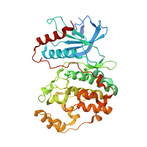
    There are a number of small-molecule inhibitors targeting the RAS/RAF/MEK/ERK signaling pathway that have either been approved or are in clinical development for oncology across a range of disease indications. The inhibition of ERK1/2 is of significant current interest, as cell lines with acquired resistance to BRAF and MEK inhibitors have been shown to maintain sensitivity to ERK1/2 inhibition in preclinical models. This article reports on our recent work to identify novel, potent, and selective reversible ERK1/2 inhibitors from a low-molecular-weight, modestly active, and highly promiscuous chemical start point, compound 4. To guide and inform the evolution of this series, inhibitor binding mode information from X-ray crystal structures was critical in the rapid exploration of this template to compound 35, which was active when tested in in vivo antitumor efficacy experiments.


  • Organizational Affiliation

    IMED Oncology and Discovery Sciences, AstraZeneca , Darwin Building, and AstraZeneca, Hodgkin Building, c/o Darwin Building, 310 Cambridge Science Park, Milton Road, Cambridge CB4 0WG, U.K.


Macromolecules
Find similar proteins by:  (by identity cutoff)  |  3D Structure
Entity ID: 1
MoleculeChains Sequence LengthOrganismDetailsImage
Mitogen-activated protein kinase 1381Homo sapiensMutation(s): 0 
Gene Names: MAPK1ERK2PRKM1PRKM2
EC: 2.7.11.24
UniProt & NIH Common Fund Data Resources
Find proteins for P28482 (Homo sapiens)
Explore P28482 
Go to UniProtKB:  P28482
PHAROS:  P28482
GTEx:  ENSG00000100030 
Entity Groups  
Sequence Clusters30% Identity50% Identity70% Identity90% Identity95% Identity100% Identity
UniProt GroupP28482
Sequence Annotations
Expand
  • Reference Sequence
Small Molecules
Ligands 2 Unique
IDChains Name / Formula / InChI Key2D Diagram3D Interactions
8XB
Query on 8XB

Download Ideal Coordinates CCD File 
C [auth A](6~{R})-5-(2-methoxyethyl)-6-methyl-2-[5-methyl-2-[(2-methylpyrazol-3-yl)amino]pyrimidin-4-yl]-6,7-dihydro-1~{H}-pyrrolo[3,2-c]pyridin-4-one
C20 H25 N7 O2
PNXHDOWDWLRBSB-CYBMUJFWSA-N
SO4
Query on SO4

Download Ideal Coordinates CCD File 
B [auth A]SULFATE ION
O4 S
QAOWNCQODCNURD-UHFFFAOYSA-L
Binding Affinity Annotations 
IDSourceBinding Affinity
8XB BindingDB:  5NHL IC50: min: 0.3, max: 130 (nM) from 3 assay(s)
Binding MOAD:  5NHL IC50: 0.3 (nM) from 1 assay(s)
Experimental Data & Validation

Experimental Data

  • Method: X-RAY DIFFRACTION
  • Resolution: 2.07 Å
  • R-Value Free: 0.237 
  • R-Value Work: 0.209 
  • R-Value Observed: 0.211 
  • Space Group: P 1 21 1
Unit Cell:
Length ( Å )Angle ( ˚ )
a = 48.91α = 90
b = 71.34β = 111.05
c = 61.16γ = 90
Software Package:
Software NamePurpose
BUSTERrefinement
xia2data reduction
Aimlessdata scaling
PHASERphasing

Structure Validation

View Full Validation Report


Ligand Structure Quality Assessment 


Entry History 

Revision History  (Full details and data files)

  • Version 1.0: 2017-04-19
    Type: Initial release
  • Version 1.1: 2017-05-10
    Changes: Database references