5NW9 | pdb_00005nw9

Crystal structure of the complex of Tdp1 with duplex DNA


Experimental Data Snapshot

  • Method: X-RAY DIFFRACTION
  • Resolution: 2.04 Å
  • R-Value Free: 
    0.271 (Depositor), 0.280 (DCC) 
  • R-Value Work: 
    0.232 (Depositor), 0.240 (DCC) 
  • R-Value Observed: 
    0.234 (Depositor) 

Starting Model: experimental
View more details

wwPDB Validation   3D Report Full Report


This is version 1.4 of the entry. See complete history


Literature

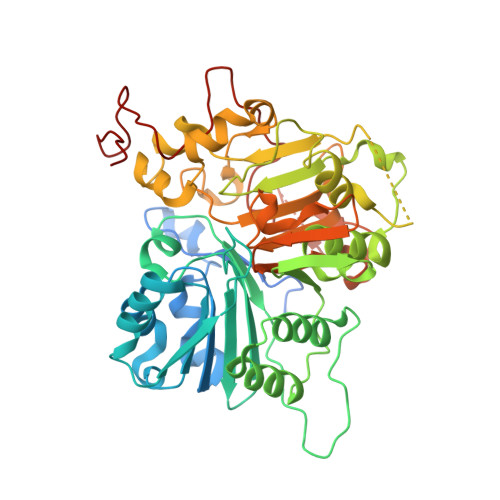
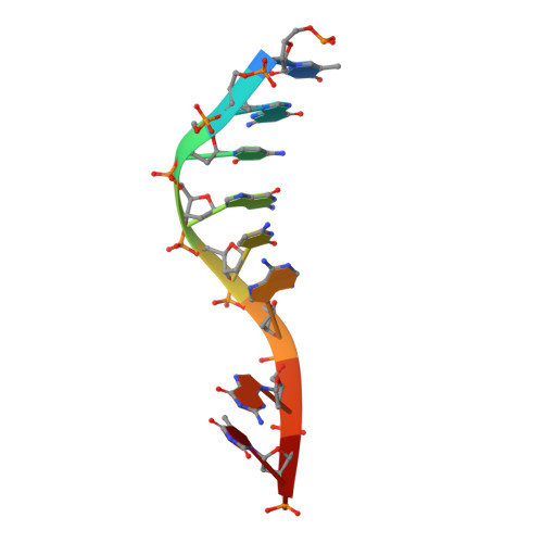
Structural basis for DNA 3'-end processing by human tyrosyl-DNA phosphodiesterase 1.

Flett, F.J.Ruksenaite, E.Armstrong, L.A.Bharati, S.Carloni, R.Morris, E.R.Mackay, C.L.Interthal, H.Richardson, J.M.

(2018) Nat Commun 9: 24-24

  • DOI: https://doi.org/10.1038/s41467-017-02530-z
  • Primary Citation of Related Structures:  
    5NW9, 5NWA

  • PubMed Abstract: 

    Tyrosyl-DNA phosphodiesterase (Tdp1) is a DNA 3'-end processing enzyme that repairs topoisomerase 1B-induced DNA damage. We use a new tool combining site-specific DNA-protein cross-linking with mass spectrometry to identify Tdp1 interactions with DNA. A conserved phenylalanine (F259) of Tdp1, required for efficient DNA processing in biochemical assays, cross-links to defined positions in DNA substrates. Crystal structures of Tdp1-DNA complexes capture the DNA repair machinery after 3'-end cleavage; these reveal how Tdp1 coordinates the 3'-phosphorylated product of nucleosidase activity and accommodates duplex DNA. A hydrophobic wedge splits the DNA ends, directing the scissile strand through a channel towards the active site. The F259 side-chain stacks against the -3 base pair, delimiting the junction of duplexed and melted DNA, and fixes the scissile strand in the channel. Our results explain why Tdp1 cleavage is non-processive and provide a molecular basis for DNA 3'-end processing by Tdp1.


  • Organizational Affiliation
    • Institute of Cell Biology, School of Biological Sciences, University of Edinburgh, The King's Buildings, Roger Land Building, Alexander Crum Brown Road, Edinburgh, EH9 3FF, UK.

Macromolecules

Find similar proteins by:  (by identity cutoff)  |  3D Structure
Entity ID: 1
MoleculeChains Sequence LengthOrganismDetailsImage
Tyrosyl-DNA phosphodiesterase 1
A, B
485Homo sapiensMutation(s): 0 
Gene Names: TDP1
EC: 3.1.4
UniProt & NIH Common Fund Data Resources
Find proteins for Q9NUW8 (Homo sapiens)
Explore Q9NUW8 
Go to UniProtKB:  Q9NUW8
PHAROS:  Q9NUW8
GTEx:  ENSG00000042088 
Entity Groups  
Sequence Clusters30% Identity50% Identity70% Identity90% Identity95% Identity100% Identity
UniProt GroupQ9NUW8
Sequence Annotations
Expand
  • Reference Sequence

Find similar nucleic acids by:  Sequence   |   3D Structure  

Entity ID: 2
MoleculeChains LengthOrganismImage
DNA (5'-D(P*TP*GP*CP*GP*CP*AP*GP*TP*A)-3')9synthetic construct
Sequence Annotations
Expand
  • Reference Sequence
Experimental Data & Validation

Experimental Data

  • Method: X-RAY DIFFRACTION
  • Resolution: 2.04 Å
  • R-Value Free:  0.271 (Depositor), 0.280 (DCC) 
  • R-Value Work:  0.232 (Depositor), 0.240 (DCC) 
  • R-Value Observed: 0.234 (Depositor) 
Space Group: P 21 21 2
Unit Cell:
Length ( Å )Angle ( ˚ )
a = 108.01α = 90
b = 195.31β = 90
c = 50.83γ = 90
Software Package:
Software NamePurpose
xia2data scaling
PHASERphasing
REFMACrefinement
PDB_EXTRACTdata extraction
PHASERphasing
xia2data reduction

Structure Validation

View Full Validation Report


Entry History & Funding Information

Deposition Data


Funding OrganizationLocationGrant Number
Biotechnology and Biological Sciences Research CouncilUnited KingdomBJ000884

Revision History  (Full details and data files)

  • Version 1.0: 2018-01-10
    Type: Initial release
  • Version 1.1: 2018-01-17
    Changes: Database references
  • Version 1.2: 2018-06-27
    Changes: Data collection, Derived calculations
  • Version 1.3: 2019-10-16
    Changes: Data collection
  • Version 1.4: 2024-01-17
    Changes: Data collection, Database references, Refinement description