5NX2

Crystal structure of thermostabilised full-length GLP-1R in complex with a truncated peptide agonist at 3.7 A resolution


Experimental Data Snapshot

  • Method: X-RAY DIFFRACTION
  • Resolution: 3.70 Å
  • R-Value Free: 
    0.334 (Depositor), 0.340 (DCC) 
  • R-Value Work: 
    0.285 (Depositor), 0.300 (DCC) 
  • R-Value Observed: 
    0.287 (Depositor) 

Starting Models: experimental
View more details

wwPDB Validation   3D Report Full Report


Ligand Structure Quality Assessment 

Created with Raphaël 2.3.0Worse 01 BetterLigand structure goodness of fit to experimental dataBest fitted SOGClick on this verticalbar to view detailsBest fitted NAGClick on this verticalbar to view details

This is version 3.1 of the entry. See complete history


Literature

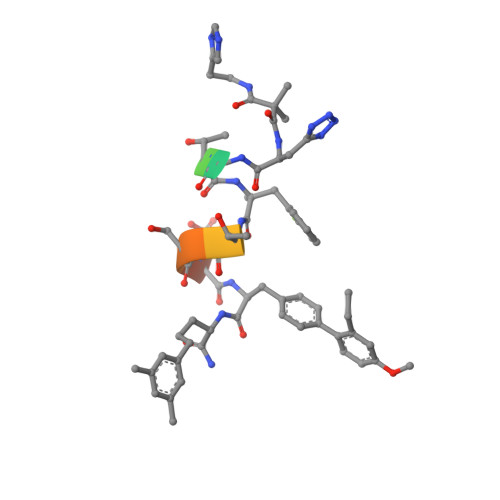
Crystal structure of the GLP-1 receptor bound to a peptide agonist.

Jazayeri, A.Rappas, M.Brown, A.J.H.Kean, J.Errey, J.C.Robertson, N.J.Fiez-Vandal, C.Andrews, S.P.Congreve, M.Bortolato, A.Mason, J.S.Baig, A.H.Teobald, I.Dore, A.S.Weir, M.Cooke, R.M.Marshall, F.H.

(2017) Nature 546: 254-258

  • DOI: https://doi.org/10.1038/nature22800
  • Primary Citation of Related Structures:  
    5NX2

  • PubMed Abstract: 

    Glucagon-like peptide 1 (GLP-1) regulates glucose homeostasis through the control of insulin release from the pancreas. GLP-1 peptide agonists are efficacious drugs for the treatment of diabetes. To gain insight into the molecular mechanism of action of GLP-1 peptides, here we report the crystal structure of the full-length GLP-1 receptor bound to a truncated peptide agonist. The peptide agonist retains an α-helical conformation as it sits deep within the receptor-binding pocket. The arrangement of the transmembrane helices reveals hallmarks of an active conformation similar to that observed in class A receptors. Guided by this structural information, we design peptide agonists with potent in vivo activity in a mouse model of diabetes.


  • Organizational Affiliation

    Heptares Therapeutics Ltd, BioPark, Broadwater Road, Welwyn Garden City, Hertfordshire AL7 3AX, UK.


Macromolecules
Find similar proteins by:  (by identity cutoff)  |  3D Structure
Entity ID: 1
MoleculeChains Sequence LengthOrganismDetailsImage
Glucagon-like peptide 1 receptor422Homo sapiensMutation(s): 12 
Gene Names: GLP1R
Membrane Entity: Yes 
UniProt & NIH Common Fund Data Resources
Find proteins for P43220 (Homo sapiens)
Explore P43220 
Go to UniProtKB:  P43220
PHAROS:  P43220
GTEx:  ENSG00000112164 
Entity Groups  
Sequence Clusters30% Identity50% Identity70% Identity90% Identity95% Identity100% Identity
UniProt GroupP43220
Glycosylation
Glycosylation Sites: 2Go to GlyGen: P43220-1
Sequence Annotations
Expand
  • Reference Sequence

Find similar proteins by:  Sequence   |   3D Structure  

Entity ID: 2
MoleculeChains Sequence LengthOrganismDetailsImage
truncated peptide agonist10synthetic constructMutation(s): 0 
Entity Groups  
Sequence Clusters30% Identity50% Identity70% Identity90% Identity95% Identity100% Identity
Sequence Annotations
Expand
  • Reference Sequence
Oligosaccharides

Help

Entity ID: 3
MoleculeChains Length2D Diagram Glycosylation3D Interactions
2-acetamido-2-deoxy-beta-D-glucopyranose-(1-4)-2-acetamido-2-deoxy-beta-D-glucopyranose
C
2N-Glycosylation
Glycosylation Resources
GlyTouCan:  G42666HT
GlyCosmos:  G42666HT
GlyGen:  G42666HT
Experimental Data & Validation

Experimental Data

  • Method: X-RAY DIFFRACTION
  • Resolution: 3.70 Å
  • R-Value Free:  0.334 (Depositor), 0.340 (DCC) 
  • R-Value Work:  0.285 (Depositor), 0.300 (DCC) 
  • R-Value Observed: 0.287 (Depositor) 
Space Group: P 31 2 1
Unit Cell:
Length ( Å )Angle ( ˚ )
a = 94.435α = 90
b = 94.435β = 90
c = 163.905γ = 120
Software Package:
Software NamePurpose
PHENIXrefinement
XDSdata reduction
Aimlessdata scaling
PHASERphasing

Structure Validation

View Full Validation Report


Ligand Structure Quality Assessment 

Created with Raphaël 2.3.0Worse 01 BetterLigand structure goodness of fit to experimental dataBest fitted SOGClick on this verticalbar to view detailsBest fitted NAGClick on this verticalbar to view details

Entry History 

Revision History  (Full details and data files)

  • Version 1.0: 2017-06-14
    Type: Initial release
  • Version 2.0: 2020-07-29
    Type: Remediation
    Reason: Carbohydrate remediation
    Changes: Atomic model, Data collection, Derived calculations, Structure summary
  • Version 3.0: 2023-11-15
    Changes: Atomic model, Data collection, Database references, Derived calculations, Structure summary
  • Version 3.1: 2024-01-17
    Changes: Refinement description