5NXS | pdb_00005nxs

Crystal Structure of Human Pro-myostatin Precursor at 4.2 A Resolution with Experimental Phases from SeMet labelling


Experimental Data Snapshot

  • Method: X-RAY DIFFRACTION
  • Resolution: 4.19 Å
  • R-Value Free: 
    0.301 (Depositor), 0.315 (DCC) 
  • R-Value Work: 
    0.274 (Depositor), 0.301 (DCC) 
  • R-Value Observed: 
    0.276 (Depositor) 

wwPDB Validation 3D Report Full Report

Validation slider image for 5NXS

This is version 1.3 of the entry. See complete history

Literature

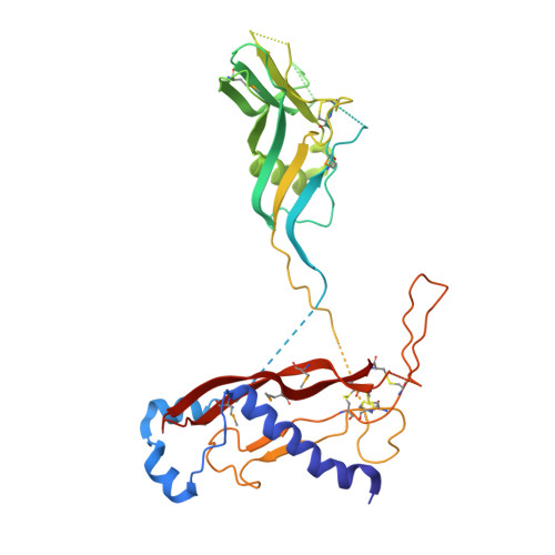
Structure of the human myostatin precursor and determinants of growth factor latency.

Cotton, T.R.Fischer, G.Wang, X.McCoy, J.C.Czepnik, M.Thompson, T.B.Hyvonen, M.

(2018) EMBO J 37: 367-383

  • DOI: https://doi.org/10.15252/embj.201797883
  • Primary Citation Related Structures: 
    5NTU, 5NXS

  • PubMed Abstract: 

    Myostatin, a key regulator of muscle mass in vertebrates, is biosynthesised as a latent precursor in muscle and is activated by sequential proteolysis of the pro-domain. To investigate the molecular mechanism by which pro-myostatin remains latent, we have determined the structure of unprocessed pro-myostatin and analysed the properties of the protein in its different forms. Crystal structures and SAXS analyses show that pro-myostatin adopts an open, V-shaped structure with a domain-swapped arrangement. The pro-mature complex, after cleavage of the furin site, has significantly reduced activity compared with the mature growth factor and persists as a stable complex that is resistant to the natural antagonist follistatin. The latency appears to be conferred by a number of distinct features that collectively stabilise the interaction of the pro-domains with the mature growth factor, enabling a regulated stepwise activation process, distinct from the prototypical pro-TGF-β1. These results provide a basis for understanding the effect of missense mutations in pro-myostatin and pave the way for the design of novel myostatin inhibitors.


  • Organizational Affiliation
    • Department of Biochemistry, University of Cambridge, Cambridge, UK.

Macromolecule Content 

  • Total Structure Weight: 77 kDa 
  • Atom Count: 3,556 
  • Modeled Residue Count: 541 
  • Deposited Residue Count: 670 
  • Unique protein chains: 1

Macromolecules

Find similar proteins by:|  3D Structure
Entity ID: 1
MoleculeChains  Sequence LengthOrganismDetailsImage
Growth/differentiation factor 8
A, B
335Homo sapiensMutation(s): 0 
Gene Names: MSTNGDF8
UniProt & NIH Common Fund Data Resources
Find proteins for O14793 (Homo sapiens)
Explore O14793 
Go to UniProtKB:  O14793
PHAROS:  O14793
GTEx:  ENSG00000138379 
Entity Groups
Sequence Clusters30% Identity50% Identity70% Identity90% Identity95% Identity100% Identity
UniProt GroupO14793
Sequence Annotations
Expand
Reference Sequence

Small Molecules

Modified Residues  1 Unique
IDChains TypeFormula2D DiagramParent
MSE
Query on MSE
A, B
L-PEPTIDE LINKINGC5 H11 N O2 SeMET

Experimental Data & Validation

Experimental Data

  • Method: X-RAY DIFFRACTION
  • Resolution: 4.19 Å
  • R-Value Free:  0.301 (Depositor), 0.315 (DCC) 
  • R-Value Work:  0.274 (Depositor), 0.301 (DCC) 
  • R-Value Observed: 0.276 (Depositor) 
Space Group: I 2 3
Unit Cell:
Length ( Å )Angle ( ˚ )
a = 196.83α = 90
b = 196.83β = 90
c = 196.83γ = 90
Software Package:
Software NamePurpose
BUSTERrefinement
Aimlessdata scaling
PDB_EXTRACTdata extraction
XDSdata reduction
AutoSolphasing

Structure Validation

View Full Validation Report


Entry History 

Deposition Data

Revision History  (Full details and data files)

  • Version 1.0: 2018-01-17
    Type: Initial release
  • Version 1.1: 2018-01-24
    Changes: Database references
  • Version 1.2: 2018-02-21
    Changes: Database references
  • Version 1.3: 2024-11-06
    Changes: Data collection, Database references, Structure summary