5O2L | pdb_00005o2l

Myosin VI motor domain in the Pre-Transition State


Experimental Data Snapshot

  • Method: X-RAY DIFFRACTION
  • Resolution: 2.20 Å
  • R-Value Free: 
    0.227 (Depositor), 0.230 (DCC) 
  • R-Value Work: 
    0.182 (Depositor) 
  • R-Value Observed: 
    0.184 (Depositor) 

Starting Model: experimental
View more details

wwPDB Validation 3D Report Full Report

Validation slider image for 5O2L

Ligand Structure Quality Assessment 


This is version 1.3 of the entry. See complete history

Literature

An intermediate along the recovery stroke of myosin VI revealed by X-ray crystallography and molecular dynamics.

Blanc, F.Isabet, T.Benisty, H.Sweeney, H.L.Cecchini, M.Houdusse, A.

(2018) Proc Natl Acad Sci U S A 115: 6213-6218

  • DOI: https://doi.org/10.1073/pnas.1711512115
  • Primary Citation Related Structures: 
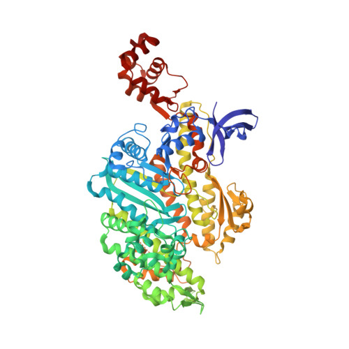
    5O2L

  • PubMed Abstract: 

    Myosins form a class of actin-based, ATPase motor proteins that mediate important cellular functions such as cargo transport and cell motility. Their functional cycle involves two large-scale swings of the lever arm: the force-generating powerstroke, which takes place on actin, and the recovery stroke during which the lever arm is reprimed into an armed configuration. Previous analyses of the prerecovery (postrigor) and postrecovery (prepowerstroke) states predicted that closure of switch II in the ATP binding site precedes the movement of the converter and the lever arm. Here, we report on a crystal structure of myosin VI, called pretransition state (PTS), which was solved at 2.2 Å resolution. Structural analysis and all-atom molecular dynamics simulations are consistent with PTS being an intermediate along the recovery stroke, where the Relay/SH1 elements adopt a postrecovery conformation, and switch II remains open. In this state, the converter appears to be largely uncoupled from the motor domain and explores an ensemble of partially reprimed configurations through extensive, reversible fluctuations. Moreover, we found that the free energy cost of hydrogen-bonding switch II to ATP is lowered by more than 10 kcal/mol compared with the prerecovery state. These results support the conclusion that closing of switch II does not initiate the recovery stroke transition in myosin VI. Rather, they suggest a mechanism in which lever arm repriming would be mostly driven by thermal fluctuations and eventually stabilized by the switch II interaction with the nucleotide in a ratchet-like fashion.


  • Organizational Affiliation
    • Structural Motility, Institut Curie, Paris Sciences et Lettres (PSL) Research University, CNRS, UMR 144, F-75005 Paris, France.

Macromolecule Content 

  • Total Structure Weight: 90.8 kDa 
  • Atom Count: 6,387 
  • Modeled Residue Count: 763 
  • Deposited Residue Count: 785 
  • Unique protein chains: 1

Macromolecules

Find similar proteins by:|  3D Structure
Entity ID: 1
MoleculeChains  Sequence LengthOrganismDetailsImage
Unconventional myosin-VI785Sus scrofaMutation(s): 0 
Gene Names: MYO6
UniProt
Find proteins for Q29122 (Sus scrofa)
Explore Q29122 
Go to UniProtKB:  Q29122
Entity Groups
Sequence Clusters30% Identity50% Identity70% Identity90% Identity95% Identity100% Identity
UniProt GroupQ29122
Sequence Annotations
Expand
Reference Sequence

Small Molecules

Ligands 4 Unique
IDChains Name / Formula / InChI Key2D Diagram3D Interactions
ADP

Query on ADP


Download:Ideal Coordinates CCD File
B [auth A]ADENOSINE-5'-DIPHOSPHATE
C10 H15 N5 O10 P2
XTWYTFMLZFPYCI-KQYNXXCUSA-N
GOL

Query on GOL


Download:Ideal Coordinates CCD File
E [auth A]
F [auth A]
G [auth A]
H [auth A]
I [auth A]
E [auth A],
F [auth A],
G [auth A],
H [auth A],
I [auth A],
J [auth A],
K [auth A],
L [auth A]
GLYCEROL
C3 H8 O3
PEDCQBHIVMGVHV-UHFFFAOYSA-N
BEF

Query on BEF


Download:Ideal Coordinates CCD File
D [auth A]BERYLLIUM TRIFLUORIDE ION
Be F3
OGIAHMCCNXDTIE-UHFFFAOYSA-K
MG

Query on MG


Download:Ideal Coordinates CCD File
C [auth A]MAGNESIUM ION
Mg
JLVVSXFLKOJNIY-UHFFFAOYSA-N

Experimental Data & Validation

Experimental Data

  • Method: X-RAY DIFFRACTION
  • Resolution: 2.20 Å
  • R-Value Free:  0.227 (Depositor), 0.230 (DCC) 
  • R-Value Work:  0.182 (Depositor) 
  • R-Value Observed: 0.184 (Depositor) 
Space Group: P 21 21 21
Unit Cell:
Length ( Å )Angle ( ˚ )
a = 72.34α = 90
b = 83.83β = 90
c = 177.66γ = 90
Software Package:
Software NamePurpose
BUSTERrefinement
Cootmodel building
XDSdata reduction
PHASERphasing

Structure Validation

View Full Validation Report


Ligand Structure Quality Assessment 


Entry History 

Deposition Data

Revision History  (Full details and data files)

  • Version 1.0: 2018-05-23
    Type: Initial release
  • Version 1.1: 2018-06-13
    Changes: Data collection, Database references
  • Version 1.2: 2018-06-20
    Changes: Data collection, Database references
  • Version 1.3: 2024-01-17
    Changes: Data collection, Database references, Derived calculations, Refinement description