5O4C

From macrocrystals to microcrystals: a strategy for membrane protein serial crystallography


Experimental Data Snapshot

  • Method: X-RAY DIFFRACTION
  • Resolution: 2.80 Å
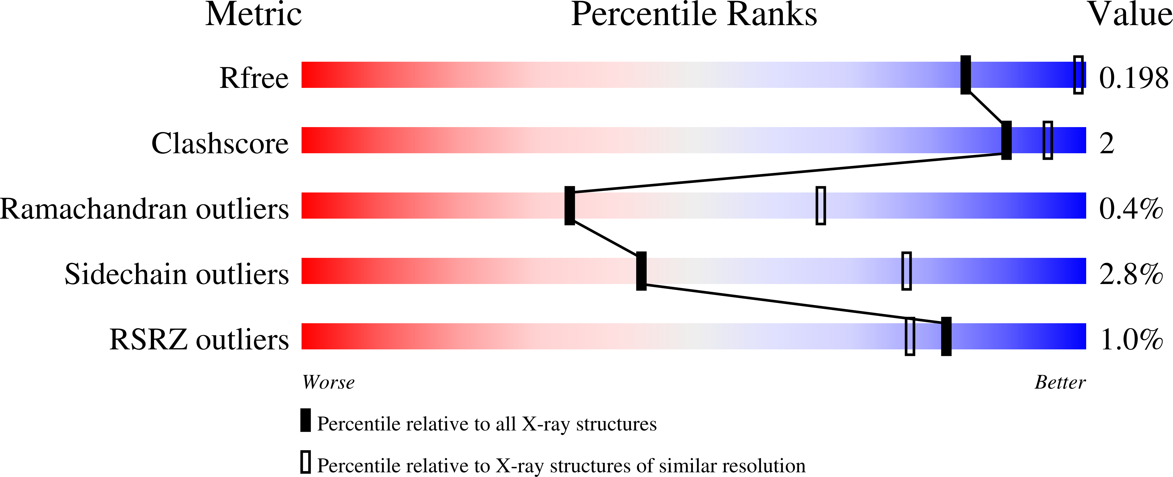
  • R-Value Free: 0.196 
  • R-Value Work: 0.165 
  • R-Value Observed: 0.167 

Starting Model: experimental
View more details

wwPDB Validation   3D Report Full Report


Ligand Structure Quality Assessment 


This is version 1.4 of the entry. See complete history


Literature

From Macrocrystals to Microcrystals: A Strategy for Membrane Protein Serial Crystallography.

Dods, R.Bath, P.Arnlund, D.Beyerlein, K.R.Nelson, G.Liang, M.Harimoorthy, R.Berntsen, P.Malmerberg, E.Johansson, L.Andersson, R.Bosman, R.Carbajo, S.Claesson, E.Conrad, C.E.Dahl, P.Hammarin, G.Hunter, M.S.Li, C.Lisova, S.Milathianaki, D.Robinson, J.Safari, C.Sharma, A.Williams, G.Wickstrand, C.Yefanov, O.Davidsson, J.DePonte, D.P.Barty, A.Branden, G.Neutze, R.

(2017) Structure 25: 1461-1468.e2

  • DOI: https://doi.org/10.1016/j.str.2017.07.002
  • Primary Citation of Related Structures:  
    5NJ4, 5O4C, 5O64

  • PubMed Abstract: 

    Serial protein crystallography was developed at X-ray free-electron lasers (XFELs) and is now also being applied at storage ring facilities. Robust strategies for the growth and optimization of microcrystals are needed to advance the field. Here we illustrate a generic strategy for recovering high-density homogeneous samples of microcrystals starting from conditions known to yield large (macro) crystals of the photosynthetic reaction center of Blastochloris viridis (RC vir ). We first crushed these crystals prior to multiple rounds of microseeding. Each cycle of microseeding facilitated improvements in the RC vir serial femtosecond crystallography (SFX) structure from 3.3-Å to 2.4-Å resolution. This approach may allow known crystallization conditions for other proteins to be adapted to exploit novel scientific opportunities created by serial crystallography.


  • Organizational Affiliation

    Department of Chemistry and Molecular Biology, University of Gothenburg, Gothenburg, Sweden.


Macromolecules
Find similar proteins by:  (by identity cutoff)  |  3D Structure
Entity ID: 1
MoleculeChains Sequence LengthOrganismDetailsImage
Photosynthetic reaction center cytochrome c subunitA [auth C]336Blastochloris viridisMutation(s): 0 
Membrane Entity: Yes 
UniProt
Find proteins for P07173 (Blastochloris viridis)
Explore P07173 
Go to UniProtKB:  P07173
Entity Groups  
Sequence Clusters30% Identity50% Identity70% Identity90% Identity95% Identity100% Identity
UniProt GroupP07173
Sequence Annotations
Expand
  • Reference Sequence
Find similar proteins by:  (by identity cutoff)  |  3D Structure
Entity ID: 2
MoleculeChains Sequence LengthOrganismDetailsImage
Reaction center protein H chainB [auth H]258Blastochloris viridisMutation(s): 0 
Membrane Entity: Yes 
UniProt
Find proteins for P06008 (Blastochloris viridis)
Explore P06008 
Go to UniProtKB:  P06008
Entity Groups  
Sequence Clusters30% Identity50% Identity70% Identity90% Identity95% Identity100% Identity
UniProt GroupP06008
Sequence Annotations
Expand
  • Reference Sequence
Find similar proteins by:  (by identity cutoff)  |  3D Structure
Entity ID: 3
MoleculeChains Sequence LengthOrganismDetailsImage
Reaction center protein L chainC [auth L]273Blastochloris viridisMutation(s): 0 
Membrane Entity: Yes 
UniProt
Find proteins for P06009 (Blastochloris viridis)
Explore P06009 
Go to UniProtKB:  P06009
Entity Groups  
Sequence Clusters30% Identity50% Identity70% Identity90% Identity95% Identity100% Identity
UniProt GroupP06009
Sequence Annotations
Expand
  • Reference Sequence
Find similar proteins by:  (by identity cutoff)  |  3D Structure
Entity ID: 4
MoleculeChains Sequence LengthOrganismDetailsImage
Reaction center protein M chainD [auth M]323Blastochloris viridisMutation(s): 0 
Membrane Entity: Yes 
UniProt
Find proteins for P06010 (Blastochloris viridis)
Explore P06010 
Go to UniProtKB:  P06010
Entity Groups  
Sequence Clusters30% Identity50% Identity70% Identity90% Identity95% Identity100% Identity
UniProt GroupP06010
Sequence Annotations
Expand
  • Reference Sequence
Small Molecules
Ligands 10 Unique
IDChains Name / Formula / InChI Key2D Diagram3D Interactions
BCB
Query on BCB

Download Ideal Coordinates CCD File 
AA [auth M],
S [auth L],
T [auth L],
Z [auth M]
BACTERIOCHLOROPHYLL B
C55 H72 Mg N4 O6
QNWPCDKNPGOYNP-DSENBSCCSA-M
BPB
Query on BPB

Download Ideal Coordinates CCD File 
BA [auth M],
U [auth L]
BACTERIOPHEOPHYTIN B
C55 H74 N4 O6
SFKCKJXMIAKQMY-GTTFDWDMSA-N
MQ7
Query on MQ7

Download Ideal Coordinates CCD File 
Y [auth M]MENAQUINONE-7
C46 H64 O2
RAKQPZMEYJZGPI-LJWNYQGCSA-N
DGA
Query on DGA

Download Ideal Coordinates CCD File 
I [auth C]DIACYL GLYCEROL
C39 H76 O5
UHUSDOQQWJGJQS-QNGWXLTQSA-N
HEC
Query on HEC

Download Ideal Coordinates CCD File 
E [auth C],
F [auth C],
G [auth C],
H [auth C]
HEME C
C34 H34 Fe N4 O4
HXQIYSLZKNYNMH-LJNAALQVSA-N
NS5
Query on NS5

Download Ideal Coordinates CCD File 
CA [auth M]15-cis-1,2-dihydroneurosporene
C40 H60
NHKJSVKSSGKUCH-DBWJSHEJSA-N
LDA
Query on LDA

Download Ideal Coordinates CCD File 
K [auth H],
P [auth H],
Q [auth H],
V [auth L]
LAURYL DIMETHYLAMINE-N-OXIDE
C14 H31 N O
SYELZBGXAIXKHU-UHFFFAOYSA-N
HTO
Query on HTO

Download Ideal Coordinates CCD File 
R [auth H],
W [auth L]
HEPTANE-1,2,3-TRIOL
C7 H16 O3
HXYCHJFUBNTKQR-RNFRBKRXSA-N
SO4
Query on SO4

Download Ideal Coordinates CCD File 
DA [auth M]
EA [auth M]
FA [auth M]
GA [auth M]
HA [auth M]
DA [auth M],
EA [auth M],
FA [auth M],
GA [auth M],
HA [auth M],
IA [auth M],
J [auth C],
JA [auth M],
L [auth H],
M [auth H],
N [auth H],
O [auth H]
SULFATE ION
O4 S
QAOWNCQODCNURD-UHFFFAOYSA-L
FE2
Query on FE2

Download Ideal Coordinates CCD File 
X [auth M]FE (II) ION
Fe
CWYNVVGOOAEACU-UHFFFAOYSA-N
Modified Residues  1 Unique
IDChains TypeFormula2D DiagramParent
FME
Query on FME
B [auth H]L-PEPTIDE LINKINGC6 H11 N O3 SMET
Experimental Data & Validation

Experimental Data

  • Method: X-RAY DIFFRACTION
  • Resolution: 2.80 Å
  • R-Value Free: 0.196 
  • R-Value Work: 0.165 
  • R-Value Observed: 0.167 
  • Space Group: P 43 21 2
Unit Cell:
Length ( Å )Angle ( ˚ )
a = 226.4α = 90
b = 226.4β = 90
c = 113.7γ = 90
Software Package:
Software NamePurpose
REFMACrefinement
CrystFELdata reduction
CrystFELdata scaling
PHASERphasing

Structure Validation

View Full Validation Report


Ligand Structure Quality Assessment 


Entry History 

Deposition Data

Revision History  (Full details and data files)

  • Version 1.0: 2017-08-16
    Type: Initial release
  • Version 1.1: 2017-09-13
    Changes: Database references
  • Version 1.2: 2018-11-14
    Changes: Data collection
  • Version 1.3: 2024-01-17
    Changes: Data collection, Database references, Derived calculations, Refinement description
  • Version 1.4: 2024-11-06
    Changes: Structure summary