5O8F

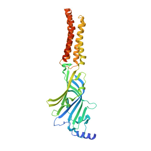
Structure of a chimaeric beta3-alpha5 GABAA receptor in complex with nanobody Nb25 and pregnanolone


Experimental Data Snapshot

  • Method: X-RAY DIFFRACTION
  • Resolution: 3.20 Å
  • R-Value Free: 0.242 
  • R-Value Work: 0.232 
  • R-Value Observed: 0.233 

Starting Model: experimental
View more details

wwPDB Validation   3D Report Full Report


Ligand Structure Quality Assessment 


This is version 2.2 of the entry. See complete history


Literature

Structural basis for GABAA receptor potentiation by neurosteroids.

Miller, P.S.Scott, S.Masiulis, S.De Colibus, L.Pardon, E.Steyaert, J.Aricescu, A.R.

(2017) Nat Struct Mol Biol 24: 986-992

  • DOI: https://doi.org/10.1038/nsmb.3484
  • Primary Citation of Related Structures:  
    5O8F, 5OJM

  • PubMed Abstract: 

    Type A γ-aminobutyric acid receptors (GABA A Rs) are the principal mediators of inhibitory neurotransmission in the human brain. Endogenous neurosteroids interact with GABA A Rs to regulate acute and chronic anxiety and are potent sedative, analgesic, anticonvulsant and anesthetic agents. Their mode of binding and mechanism of receptor potentiation, however, remain unknown. Here we report crystal structures of a chimeric GABA A R construct in apo and pregnanolone-bound states. The neurosteroid-binding site is mechanically coupled to the helices lining the ion channel pore and modulates the desensitization-gate conformation. We demonstrate that the equivalent site is responsible for physiological, heteromeric GABA A R potentiation and explain the contrasting modulatory properties of 3a versus 3b neurosteroid epimers. These results illustrate how peripheral lipid ligands can regulate the desensitization gate of GABA A Rs, a process of broad relevance to pentameric ligand-gated ion channels.


  • Organizational Affiliation

    Division of Structural Biology, Wellcome Trust Centre for Human Genetics, University of Oxford, Oxford, UK.


Macromolecules
Find similar proteins by:  (by identity cutoff)  |  3D Structure
Entity ID: 1
MoleculeChains Sequence LengthOrganismDetailsImage
Gamma-aminobutyric acid receptor subunit beta-3,Gamma-aminobutyric acid receptor subunit alpha-5,Gamma-aminobutyric acid receptor subunit alpha-5
A, B, C, D, E
367Homo sapiensMutation(s): 0 
Gene Names: GABRB3GABRA5
Membrane Entity: Yes 
UniProt & NIH Common Fund Data Resources
Find proteins for P31644 (Homo sapiens)
Explore P31644 
Go to UniProtKB:  P31644
PHAROS:  P31644
GTEx:  ENSG00000186297 
Find proteins for P28472 (Homo sapiens)
Explore P28472 
Go to UniProtKB:  P28472
PHAROS:  P28472
GTEx:  ENSG00000166206 
Entity Groups  
Sequence Clusters30% Identity50% Identity70% Identity90% Identity95% Identity100% Identity
UniProt GroupsP31644P28472
Glycosylation
Glycosylation Sites: 2Go to GlyGen: P28472-1P31644-1
Sequence Annotations
Expand
  • Reference Sequence
Find similar proteins by:  (by identity cutoff)  |  3D Structure
Entity ID: 2
MoleculeChains Sequence LengthOrganismDetailsImage
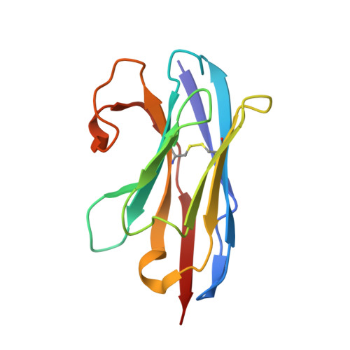
Nanobody Nb25F [auth K],
J [auth O]
125Lama glamaMutation(s): 0 
Entity Groups  
Sequence Clusters30% Identity50% Identity70% Identity90% Identity95% Identity100% Identity
Sequence Annotations
Expand
  • Reference Sequence
Find similar proteins by:  (by identity cutoff)  |  3D Structure
Entity ID: 3
MoleculeChains Sequence LengthOrganismDetailsImage
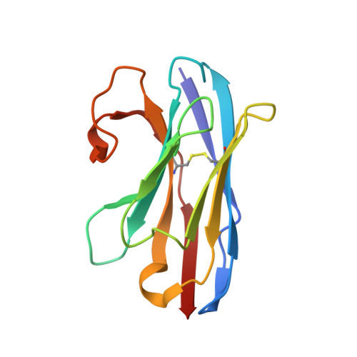
Nanobody Nb25G [auth L],
H [auth M],
I [auth N]
124Lama glamaMutation(s): 0 
Entity Groups  
Sequence Clusters30% Identity50% Identity70% Identity90% Identity95% Identity100% Identity
Sequence Annotations
Expand
  • Reference Sequence
Oligosaccharides

Help

Entity ID: 4
MoleculeChains Length2D Diagram Glycosylation3D Interactions
alpha-D-mannopyranose-(1-2)-alpha-D-mannopyranose-(1-3)-alpha-D-mannopyranose-(1-6)-[alpha-D-mannopyranose-(1-3)]beta-D-mannopyranose-(1-4)-2-acetamido-2-deoxy-beta-D-glucopyranose-(1-4)-2-acetamido-2-deoxy-beta-D-glucopyranoseK [auth F],
L [auth G],
M [auth H],
N [auth I],
O [auth J]
7N-Glycosylation
Glycosylation Resources
GlyTouCan:  G16404YH
GlyCosmos:  G16404YH
GlyGen:  G16404YH
Small Molecules
Ligands 2 Unique
IDChains Name / Formula / InChI Key2D Diagram3D Interactions
P9N
Query on P9N

Download Ideal Coordinates CCD File 
R [auth B],
T [auth C],
V [auth D],
W [auth E],
X [auth E]
Pregnanolone
C21 H34 O2
AURFZBICLPNKBZ-YZRLXODZSA-N
NAG
Query on NAG

Download Ideal Coordinates CCD File 
P [auth A],
Q [auth B],
S [auth C],
U [auth D],
Y [auth E]
2-acetamido-2-deoxy-beta-D-glucopyranose
C8 H15 N O6
OVRNDRQMDRJTHS-FMDGEEDCSA-N
Experimental Data & Validation

Experimental Data

  • Method: X-RAY DIFFRACTION
  • Resolution: 3.20 Å
  • R-Value Free: 0.242 
  • R-Value Work: 0.232 
  • R-Value Observed: 0.233 
  • Space Group: I 2 3
Unit Cell:
Length ( Å )Angle ( ˚ )
a = 290.102α = 90
b = 290.102β = 90
c = 290.102γ = 90
Software Package:
Software NamePurpose
REFMACrefinement
HKL-2000data reduction
Aimlessdata scaling
PHASERphasing
Cootmodel building

Structure Validation

View Full Validation Report


Ligand Structure Quality Assessment 


Entry History & Funding Information

Deposition Data


Funding OrganizationLocationGrant Number
Biotechnology and Biological Sciences Research CouncilUnited KingdomBB/M024709/1
Medical Research Council (United Kingdom)United KingdomMR/L009609/1
Medical Research Council (United Kingdom)United KingdomMC_UP_1201/15
Wellcome TrustUnited Kingdom105247/Z/14/Z
Human Frontier Science ProgramFranceRGP0065/2014
Wellcome TrustUnited Kingdom090532/Z/09/Z

Revision History  (Full details and data files)

  • Version 1.0: 2017-11-01
    Type: Initial release
  • Version 1.1: 2017-11-15
    Changes: Database references
  • Version 2.0: 2020-07-29
    Type: Remediation
    Reason: Carbohydrate remediation
    Changes: Atomic model, Data collection, Derived calculations, Structure summary
  • Version 2.1: 2024-01-17
    Changes: Data collection, Database references, Derived calculations, Refinement description, Structure summary
  • Version 2.2: 2024-11-06
    Changes: Structure summary