5O8V | pdb_00005o8v

Covalent Inhibitor 4a bound to the Lipid Pocket of p38alpha Mutant S251C


Experimental Data Snapshot

  • Method: X-RAY DIFFRACTION
  • Resolution: 2.00 Å
  • R-Value Free: 
    0.269 (Depositor), 0.271 (DCC) 
  • R-Value Work: 
    0.206 (Depositor), 0.214 (DCC) 
  • R-Value Observed: 
    0.209 (Depositor) 

Starting Model: experimental
View more details

wwPDB Validation 3D Report Full Report

Validation slider image for 5O8V

Ligand Structure Quality Assessment 


This is version 1.4 of the entry. See complete history

Literature

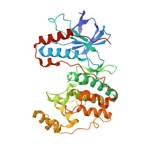
Covalent Lipid Pocket Ligands Targeting p38 alpha MAPK Mutants.

Buhrmann, M.Hardick, J.Weisner, J.Quambusch, L.Rauh, D.

(2017) Angew Chem Int Ed Engl 56: 13232-13236

  • DOI: https://doi.org/10.1002/anie.201706345
  • Primary Citation Related Structures: 
    5O8U, 5O8V

  • PubMed Abstract: 

    A chemical genetic approach is presented to covalently target a unique lipid binding pocket in the protein kinase p38α, whose function is not yet known. Based on a series of cocrystal structures, a library of 2-arylquinazolines that were decorated with electrophiles were designed and synthesized to covalently target tailored p38α mutants containing artificially introduced cysteine residues. Matching protein-ligand pairs were identified by MS analysis and further validated by MS/MS studies and protein crystallography. The covalent ligands that emerged from this approach showed excellent selectivity towards a single p38α mutant and will be applicable as suitable probes in future studies of biological systems to dissect the function of the lipid pocket by means of pharmacological perturbations.


  • Organizational Affiliation
    • Faculty of Chemistry and Chemical Biology, TU Dortmund University, Otto-Hahn-Strasse 4a, 44227, Dortmund, Germany.

Macromolecule Content 

  • Total Structure Weight: 41.77 kDa 
  • Atom Count: 2,708 
  • Modeled Residue Count: 328 
  • Deposited Residue Count: 360 
  • Unique protein chains: 1

Macromolecules

Find similar proteins by:|  3D Structure
Entity ID: 1
MoleculeChains  Sequence LengthOrganismDetailsImage
Mitogen-activated protein kinase 14360Homo sapiensMutation(s): 1 
Gene Names: MAPK14CSBPCSBP1CSBP2CSPB1MXI2SAPK2A
EC: 2.7.11.24
UniProt & NIH Common Fund Data Resources
Find proteins for Q16539 (Homo sapiens)
Explore Q16539 
Go to UniProtKB:  Q16539
PHAROS:  Q16539
GTEx:  ENSG00000112062 
Entity Groups
Sequence Clusters30% Identity50% Identity70% Identity90% Identity95% Identity100% Identity
UniProt GroupQ16539
Sequence Annotations
Expand
Reference Sequence

Small Molecules

Ligands 1 Unique
IDChains Name / Formula / InChI Key2D Diagram3D Interactions
9O2

Query on 9O2


Download:Ideal Coordinates CCD File
B [auth A]~{N}-[3-[7-azanyl-4-(2-phenylethylamino)quinazolin-2-yl]phenyl]propanamide
C25 H25 N5 O
VKNJKOQEMPCERA-UHFFFAOYSA-N

Experimental Data & Validation

Experimental Data

  • Method: X-RAY DIFFRACTION
  • Resolution: 2.00 Å
  • R-Value Free:  0.269 (Depositor), 0.271 (DCC) 
  • R-Value Work:  0.206 (Depositor), 0.214 (DCC) 
  • R-Value Observed: 0.209 (Depositor) 
Space Group: P 21 21 21
Unit Cell:
Length ( Å )Angle ( ˚ )
a = 65.2α = 90
b = 74.25β = 90
c = 77.82γ = 90
Software Package:
Software NamePurpose
REFMACrefinement
XDSdata reduction
XSCALEdata scaling
PHASERphasing

Structure Validation

View Full Validation Report


Ligand Structure Quality Assessment 


Entry History 

Deposition Data

Revision History  (Full details and data files)

  • Version 1.0: 2017-09-06
    Type: Initial release
  • Version 1.1: 2017-10-25
    Changes: Database references
  • Version 1.2: 2018-05-02
    Changes: Data collection
  • Version 1.3: 2024-01-17
    Changes: Data collection, Database references, Refinement description
  • Version 1.4: 2024-10-23
    Changes: Structure summary