


Structural insights into collagen-binding by platelet receptor Glycoprotein VI
Feitsma, L.J., Brondijk, T.H.C., Jarvis, G., Hagemans, D., Bihan, D., Jerah, N., Versteeg, M., Farndale, R.W., Huizinga, E.G.To be published.
Experimental Data Snapshot
Starting Model: experimental
View more details
Entity ID: 1 | |||||
|---|---|---|---|---|---|
| Molecule | Chains | Sequence Length | Organism | Details | Image |
| Platelet glycoprotein VI | 181 | Homo sapiens | Mutation(s): 0 Gene Names: GP6 | 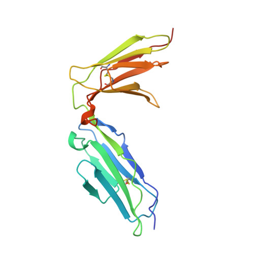 | |
UniProt & NIH Common Fund Data Resources | |||||
PHAROS: Q9HCN6 GTEx: ENSG00000088053 | |||||
Entity Groups | |||||
| Sequence Clusters | 30% Identity50% Identity70% Identity90% Identity95% Identity100% Identity | ||||
| UniProt Group | Q9HCN6 | ||||
Glycosylation | |||||
| Glycosylation Sites: 1 | Go to GlyGen: Q9HCN6-1 | ||||
Sequence AnnotationsExpand | |||||
Reference Sequence | |||||
| Ligands 4 Unique | |||||
|---|---|---|---|---|---|
| ID | Chains | Name / Formula / InChI Key | 2D Diagram | 3D Interactions | |
| PG6 Download:Ideal Coordinates CCD File | K [auth A], S [auth B] | 1-(2-METHOXY-ETHOXY)-2-{2-[2-(2-METHOXY-ETHOXY]-ETHOXY}-ETHANE C12 H26 O6 DMDPGPKXQDIQQG-UHFFFAOYSA-N |  | ||
| NAG Download:Ideal Coordinates CCD File | L [auth A], T [auth B], W [auth C], Z [auth D] | 2-acetamido-2-deoxy-beta-D-glucopyranose C8 H15 N O6 OVRNDRQMDRJTHS-FMDGEEDCSA-N |  | ||
| PO4 Download:Ideal Coordinates CCD File | J [auth A], R [auth B] | PHOSPHATE ION O4 P NBIIXXVUZAFLBC-UHFFFAOYSA-K |  | ||
| CL Download:Ideal Coordinates CCD File | E [auth A] F [auth A] G [auth A] H [auth A] I [auth A] | CHLORIDE ION Cl VEXZGXHMUGYJMC-UHFFFAOYSA-M |  | ||
| Length ( Å ) | Angle ( ˚ ) |
|---|---|
| a = 78.65 | α = 90 |
| b = 44.05 | β = 104.69 |
| c = 117.66 | γ = 90 |
| Software Name | Purpose |
|---|---|
| REFMAC | refinement |
| iMOSFLM | data reduction |
| Aimless | data scaling |
| PHASER | phasing |
| Funding Organization | Location | Grant Number |
|---|---|---|
| Netherlands Organization for Scientific Research | Netherlands | ECHO 700.58.006 |


