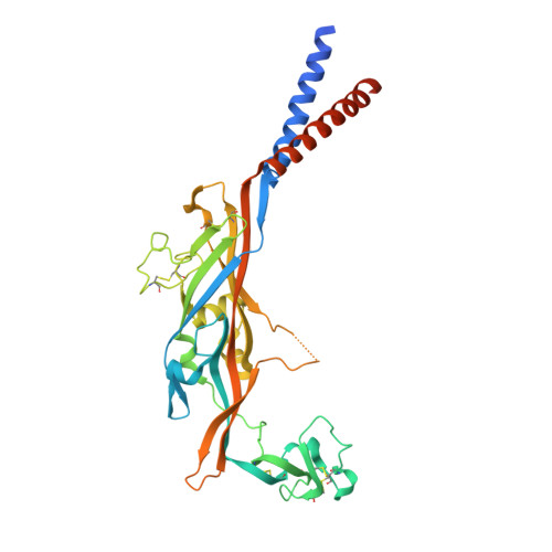
5SVJ

Crystal structure of the ATP-gated human P2X3 ion channel in the closed, apo state


Experimental Data Snapshot

  • Method: X-RAY DIFFRACTION
  • Resolution: 2.98 Å
  • R-Value Free: 0.259 
  • R-Value Work: 0.212 
  • R-Value Observed: 0.215 

wwPDB Validation   3D Report Full Report


Ligand Structure Quality Assessment 


This is version 1.5 of the entry. See complete history


Literature

X-ray structures define human P2X3 receptor gating cycle and antagonist action.

Mansoor, S.E.Lu, W.Oosterheert, W.Shekhar, M.Tajkhorshid, E.Gouaux, E.

(2016) Nature 538: 66-71

  • DOI: https://doi.org/10.1038/nature19367
  • Primary Citation of Related Structures:  
    5SVJ, 5SVK, 5SVL, 5SVM, 5SVP, 5SVQ, 5SVR, 5SVS, 5SVT

  • PubMed Abstract: 

    P2X receptors are trimeric, non-selective cation channels activated by ATP that have important roles in the cardiovascular, neuronal and immune systems. Despite their central function in human physiology and although they are potential targets of therapeutic agents, there are no structures of human P2X receptors. The mechanisms of receptor desensitization and ion permeation, principles of antagonism, and complete structures of the pore-forming transmembrane domains of these receptors remain unclear. Here we report X-ray crystal structures of the human P2X 3 receptor in apo/resting, agonist-bound/open-pore, agonist-bound/closed-pore/desensitized and antagonist-bound/closed states. The open state structure harbours an intracellular motif we term the 'cytoplasmic cap', which stabilizes the open state of the ion channel pore and creates lateral, phospholipid-lined cytoplasmic fenestrations for water and ion egress. The competitive antagonists TNP-ATP and A-317491 stabilize the apo/resting state and reveal the interactions responsible for competitive inhibition. These structures illuminate the conformational rearrangements that underlie P2X receptor gating and provide a foundation for the development of new pharmacological agents.


  • Organizational Affiliation

    Vollum Institute, Oregon Health &Science University, Portland, Oregon 97239, USA.


Macromolecules
Find similar proteins by:  (by identity cutoff)  |  3D Structure
Entity ID: 1
MoleculeChains Sequence LengthOrganismDetailsImage
P2X purinoceptor 3363Homo sapiensMutation(s): 3 
Gene Names: P2RX3
Membrane Entity: Yes 
UniProt & NIH Common Fund Data Resources
Find proteins for P56373 (Homo sapiens)
Explore P56373 
Go to UniProtKB:  P56373
PHAROS:  P56373
GTEx:  ENSG00000109991 
Entity Groups  
Sequence Clusters30% Identity50% Identity70% Identity90% Identity95% Identity100% Identity
UniProt GroupP56373
Glycosylation
Glycosylation Sites: 3Go to GlyGen: P56373-1
Sequence Annotations
Expand
  • Reference Sequence
Small Molecules
Ligands 5 Unique
IDChains Name / Formula / InChI Key2D Diagram3D Interactions
NAG
Query on NAG

Download Ideal Coordinates CCD File 
B [auth A],
C [auth A],
D [auth A]
2-acetamido-2-deoxy-beta-D-glucopyranose
C8 H15 N O6
OVRNDRQMDRJTHS-FMDGEEDCSA-N
PG4
Query on PG4

Download Ideal Coordinates CCD File 
G [auth A]TETRAETHYLENE GLYCOL
C8 H18 O5
UWHCKJMYHZGTIT-UHFFFAOYSA-N
EDO
Query on EDO

Download Ideal Coordinates CCD File 
H [auth A]1,2-ETHANEDIOL
C2 H6 O2
LYCAIKOWRPUZTN-UHFFFAOYSA-N
MG
Query on MG

Download Ideal Coordinates CCD File 
E [auth A]MAGNESIUM ION
Mg
JLVVSXFLKOJNIY-UHFFFAOYSA-N
NA
Query on NA

Download Ideal Coordinates CCD File 
F [auth A]SODIUM ION
Na
FKNQFGJONOIPTF-UHFFFAOYSA-N
Experimental Data & Validation

Experimental Data

  • Method: X-RAY DIFFRACTION
  • Resolution: 2.98 Å
  • R-Value Free: 0.259 
  • R-Value Work: 0.212 
  • R-Value Observed: 0.215 
  • Space Group: H 3 2
Unit Cell:
Length ( Å )Angle ( ˚ )
a = 120.17α = 90
b = 120.17β = 90
c = 236.58γ = 120
Software Package:
Software NamePurpose
PHENIXrefinement
XDSdata reduction
XDSdata scaling
PHENIXphasing

Structure Validation

View Full Validation Report


Ligand Structure Quality Assessment 


Entry History & Funding Information

Deposition Data


Funding OrganizationLocationGrant Number
National Institutes of Health/National Institute of General Medical Sciences (NIH/NIGMS)United StatesR01GM100400
National Institutes of Health/National Institute of General Medical Sciences (NIH/NIGMS)United States5F32GM108391

Revision History  (Full details and data files)

  • Version 1.0: 2016-09-28
    Type: Initial release
  • Version 1.1: 2016-10-19
    Changes: Database references
  • Version 1.2: 2017-09-20
    Changes: Author supporting evidence, Database references, Derived calculations
  • Version 1.3: 2019-12-25
    Changes: Author supporting evidence
  • Version 1.4: 2020-07-29
    Type: Remediation
    Reason: Carbohydrate remediation
    Changes: Data collection, Derived calculations, Structure summary
  • Version 1.5: 2024-10-30
    Changes: Data collection, Database references, Structure summary