5T48 | pdb_00005t48

Crystal structure of the D. melanogaster eIF4E-eIF4G complex without lateral contact


Experimental Data Snapshot

  • Method: X-RAY DIFFRACTION
  • Resolution: 2.19 Å
  • R-Value Free: 
    0.231 (Depositor), 0.238 (DCC) 
  • R-Value Work: 
    0.208 (Depositor), 0.216 (DCC) 
  • R-Value Observed: 
    0.210 (Depositor) 

Starting Model: experimental
View more details

wwPDB Validation 3D Report Full Report

Validation slider image for 5T48

Ligand Structure Quality Assessment 


This is version 1.3 of the entry. See complete history

Literature

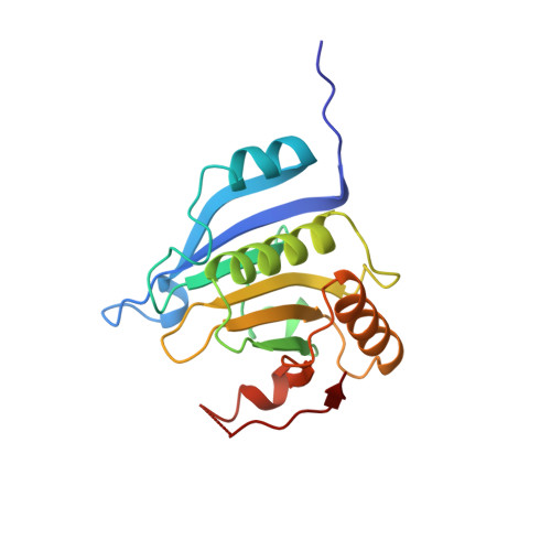
The Structures of eIF4E-eIF4G Complexes Reveal an Extended Interface to Regulate Translation Initiation.

Gruner, S.Peter, D.Weber, R.Wohlbold, L.Chung, M.Y.Weichenrieder, O.Valkov, E.Igreja, C.Izaurralde, E.

(2016) Mol Cell 64: 467-479

  • DOI: https://doi.org/10.1016/j.molcel.2016.09.020
  • Primary Citation Related Structures: 
    5T46, 5T47, 5T48

  • PubMed Abstract: 

    Eukaryotic initiation factor 4G (eIF4G) plays a central role in translation initiation through its interactions with the cap-binding protein eIF4E. This interaction is a major drug target for repressing translation and is naturally regulated by 4E-binding proteins (4E-BPs). 4E-BPs and eIF4G compete for binding to the eIF4E dorsal surface via a shared canonical 4E-binding motif, but also contain auxiliary eIF4E-binding sequences, which were assumed to contact non-overlapping eIF4E surfaces. However, it is unknown how metazoan eIF4G auxiliary sequences bind eIF4E. Here, we describe crystal structures of human and Drosophila melanogaster eIF4E-eIF4G complexes, which unexpectedly reveal that the eIF4G auxiliary sequences bind to the lateral surface of eIF4E, using a similar mode to that of 4E-BPs. Our studies provide a molecular model of the eIF4E-eIF4G complex, shed light on the competition mechanism of 4E-BPs, and enable the rational design of selective eIF4G inhibitors to dampen dysregulated translation in disease.


  • Organizational Affiliation
    • Department of Biochemistry, Max Planck Institute for Developmental Biology, Spemannstrasse 35, 72076 Tübingen, Germany.

Macromolecule Content 

  • Total Structure Weight: 29.2 kDa 
  • Atom Count: 1,851 
  • Modeled Residue Count: 217 
  • Deposited Residue Count: 248 
  • Unique protein chains: 2

Macromolecules

Find similar proteins by:|  3D Structure
Entity ID: 1
MoleculeChains  Sequence LengthOrganismDetailsImage
Eukaryotic translation initiation factor 4E184Drosophila melanogasterMutation(s): 0 
Gene Names: eIF-4EEif4eEIF4FCG4035
UniProt
Find proteins for P48598 (Drosophila melanogaster)
Explore P48598 
Go to UniProtKB:  P48598
Entity Groups
Sequence Clusters30% Identity50% Identity70% Identity90% Identity95% Identity100% Identity
UniProt GroupP48598
Sequence Annotations
Expand
Reference Sequence
Find similar proteins by:|  3D Structure
Entity ID: 2
MoleculeChains  Sequence LengthOrganismDetailsImage
Eukaryotic translation initiation factor 4G, isoform A64Drosophila melanogasterMutation(s): 0 
Gene Names: eIF4GeIF-4GeIF4G-RACG10811Dmel_CG10811
UniProt
Find proteins for O61380 (Drosophila melanogaster)
Explore O61380 
Go to UniProtKB:  O61380
Entity Groups
Sequence Clusters30% Identity50% Identity70% Identity90% Identity95% Identity100% Identity
UniProt GroupO61380
Sequence Annotations
Expand
Reference Sequence

Small Molecules

Ligands 1 Unique
IDChains Name / Formula / InChI Key2D Diagram3D Interactions
MGP

Query on MGP


Download:Ideal Coordinates CCD File
C [auth A]7-METHYL-GUANOSINE-5'-TRIPHOSPHATE
C11 H19 N5 O14 P3
DKVRNHPCAOHRSI-KQYNXXCUSA-O

Experimental Data & Validation

Experimental Data

  • Method: X-RAY DIFFRACTION
  • Resolution: 2.19 Å
  • R-Value Free:  0.231 (Depositor), 0.238 (DCC) 
  • R-Value Work:  0.208 (Depositor), 0.216 (DCC) 
  • R-Value Observed: 0.210 (Depositor) 
Space Group: P 21 21 21
Unit Cell:
Length ( Å )Angle ( ˚ )
a = 45.622α = 90
b = 66.196β = 90
c = 75.618γ = 90
Software Package:
Software NamePurpose
PHENIXrefinement
XDSdata reduction
Aimlessdata scaling
PHASERphasing

Structure Validation

View Full Validation Report


Ligand Structure Quality Assessment 


Entry History 

Deposition Data

Revision History  (Full details and data files)

  • Version 1.0: 2016-10-26
    Type: Initial release
  • Version 1.1: 2016-11-02
    Changes: Database references
  • Version 1.2: 2016-11-16
    Changes: Database references
  • Version 1.3: 2024-01-17
    Changes: Data collection, Database references, Refinement description