5T4H | pdb_00005t4h

Human DPP4 in complex with ligand 34n


Experimental Data Snapshot

  • Method: X-RAY DIFFRACTION
  • Resolution: 2.61 Å
  • R-Value Free: 
    0.226 (Depositor), 0.227 (DCC) 
  • R-Value Work: 
    0.148 (Depositor), 0.151 (DCC) 
  • R-Value Observed: 
    0.152 (Depositor) 

Starting Model: experimental
View more details

wwPDB Validation 3D Report Full Report

Validation slider image for 5T4H

Ligand Structure Quality Assessment 


This is version 2.2 of the entry. See complete history

Literature

Scaffold-hopping from xanthines to tricyclic guanines: A case study of dipeptidyl peptidase 4 (DPP4) inhibitors.

Pissarnitski, D.A.Zhao, Z.Cole, D.Wu, W.L.Domalski, M.Clader, J.W.Scapin, G.Voigt, J.Soriano, A.Kelly, T.Powles, M.A.Yao, Z.Burnett, D.A.

(2016) Bioorg Med Chem 24: 5534-5545

  • DOI: https://doi.org/10.1016/j.bmc.2016.09.007
  • Primary Citation Related Structures: 
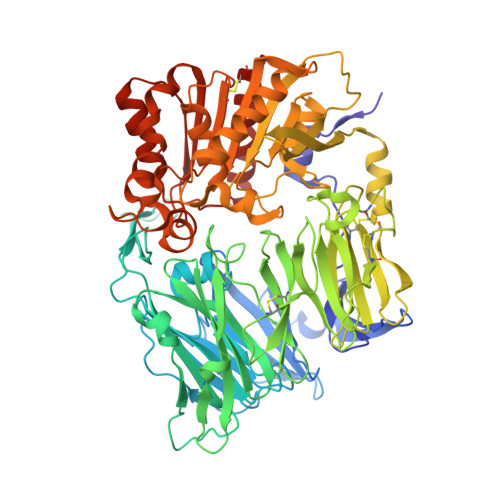
    5T4B, 5T4E, 5T4F, 5T4H

  • PubMed Abstract: 

    Molecular modeling of unbound tricyclic guanine scaffolds indicated that they can serve as effective bioisosteric replacements of xanthines. This notion was further confirmed by a combination of X-ray crystallography and SAR studies, indicating that tricyclic guanine DPP4 inhibitors mimic the binding mode of xanthine inhibitors, exemplified by linagliptin. Realization of the bioisosteric relationship between these scaffolds potentially will lead to a wider application of cyclic guanines as xanthine replacements in drug discovery programs for a variety of biological targets. Newly designed DPP4 inhibitors achieved sub-nanomolar potency range and demonstrated oral activity in vivo in mouse glucose tolerance test.


  • Organizational Affiliation
    • Department of Medicinal Chemistry, Merck Research Laboratories, 2000 Galloping Hill Rd., Kenilworth, NJ 07033, United States.

Macromolecule Content 

  • Total Structure Weight: 173.93 kDa 
  • Atom Count: 13,686 
  • Modeled Residue Count: 1,456 
  • Deposited Residue Count: 1,456 
  • Unique protein chains: 1

Macromolecules

Find similar proteins by:|  3D Structure
Entity ID: 1
MoleculeChains  Sequence LengthOrganismDetailsImage
Dipeptidyl peptidase 4
A, B
728Homo sapiensMutation(s): 0 
Gene Names: DPP4ADCP2CD26
EC: 3.4.14.5
UniProt & NIH Common Fund Data Resources
Find proteins for P27487 (Homo sapiens)
Explore P27487 
Go to UniProtKB:  P27487
PHAROS:  P27487
GTEx:  ENSG00000197635 
Entity Groups
Sequence Clusters30% Identity50% Identity70% Identity90% Identity95% Identity100% Identity
UniProt GroupP27487
Glycosylation
Glycosylation Sites: 6Go to GlyGen: P27487-1
Sequence Annotations
Expand
Reference Sequence

Oligosaccharides

Help  
Entity ID: 2
MoleculeChains Length2D Diagram GlycosylationD Interactions
2-acetamido-2-deoxy-beta-D-glucopyranose-(1-4)-2-acetamido-2-deoxy-beta-D-glucopyranose
C, D, E, F, G
C, D, E, F, G, H, I
2N-Glycosylation
Glycosylation Resources
GlyTouCan: G42666HT
GlyCosmos: G42666HT
GlyGen: G42666HT

Small Molecules

Ligands 3 Unique
IDChains Name / Formula / InChI Key2D Diagram3D Interactions
75J

Query on 75J


Download:Ideal Coordinates CCD File
M [auth A],
Q [auth B]
2-({2-[(3R)-3-aminopiperidin-1-yl]-3-(but-2-yn-1-yl)-4-oxo-3,4-dihydro-5H-imidazo[2,1-b]purin-5-yl}methyl)benzonitrile
C24 H24 N8 O
FEXBDHCWKVFWPP-LJQANCHMSA-N
NAG

Query on NAG


Download:Ideal Coordinates CCD File
J [auth A],
K [auth A],
L [auth A],
O [auth B],
P [auth B]
2-acetamido-2-deoxy-beta-D-glucopyranose
C8 H15 N O6
OVRNDRQMDRJTHS-FMDGEEDCSA-N
NA

Query on NA


Download:Ideal Coordinates CCD File
N [auth A]SODIUM ION
Na
FKNQFGJONOIPTF-UHFFFAOYSA-N

Experimental Data & Validation

Experimental Data

  • Method: X-RAY DIFFRACTION
  • Resolution: 2.61 Å
  • R-Value Free:  0.226 (Depositor), 0.227 (DCC) 
  • R-Value Work:  0.148 (Depositor), 0.151 (DCC) 
  • R-Value Observed: 0.152 (Depositor) 
Space Group: P 21 21 21
Unit Cell:
Length ( Å )Angle ( ˚ )
a = 118.125α = 90
b = 126.008β = 90
c = 137.546γ = 90
Software Package:
Software NamePurpose
BUSTERrefinement
SCALAdata scaling
PDB_EXTRACTdata extraction
XDSdata reduction
REFMACphasing

Structure Validation

View Full Validation Report


Ligand Structure Quality Assessment 


Entry History 

Deposition Data

  • Released Date: 2016-10-05 
  • Deposition Author(s): Scapin, G.

Revision History  (Full details and data files)

  • Version 1.0: 2016-10-05
    Type: Initial release
  • Version 1.1: 2016-10-26
    Changes: Database references
  • Version 2.0: 2020-07-29
    Type: Remediation
    Reason: Carbohydrate remediation
    Changes: Advisory, Atomic model, Data collection, Database references, Derived calculations, Structure summary
  • Version 2.1: 2023-10-04
    Changes: Data collection, Database references, Refinement description, Structure summary
  • Version 2.2: 2024-10-16
    Changes: Structure summary