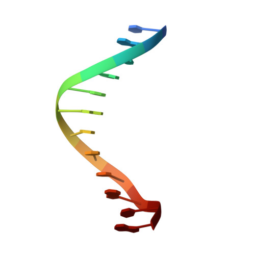
5T4W

Crystal structure of a DNA sequence d (CGTGAATTCACG) with DAPI


Experimental Data Snapshot

  • Method: X-RAY DIFFRACTION
  • Resolution: 2.30 Å
  • R-Value Free: 0.258 
  • R-Value Work: 0.216 
  • R-Value Observed: 0.220 

Starting Model: experimental
View more details

wwPDB Validation   3D Report Full Report


Ligand Structure Quality Assessment 


This is version 1.3 of the entry. See complete history


Literature

Crystal structure of the DNA sequence d(CGTGAATTCACG)2 with DAPI.

Sbirkova-Dimitrova, H.I.Shivachev, B.

(2017) Acta Crystallogr F Struct Biol Commun 73: 500-504

  • DOI: https://doi.org/10.1107/S2053230X17011384
  • Primary Citation of Related Structures:  
    5T4W

  • PubMed Abstract: 

    The structure of 4',6-diamidine-2-phenylindole (DAPI) bound to the synthetic B-DNA oligonucleotide d(CGTGAATTCACG) has been solved in space group P2 1 2 1 2 1 by single-crystal X-ray diffraction at a resolution of 2.2 Å. The structure is nearly isomorphous to that of the previously reported crystal structure of the oligonucleotide d(CGTGAATTCACG) alone. The adjustments in crystal packing between the native DNA molecule and the DNA-DAPI complex are described. DAPI lies in the narrow minor groove near the centre of the B-DNA fragment, positioned over the A-T base pairs. It is bound to the DNA by hydrogen-bonding and van der Waals interactions. Comparison of the two structures (with and without ligand) shows that DAPI inserts into the minor groove, displacing the ordered spine waters. Indeed, as DAPI is hydrophobic it confers this behaviour on the DNA and thus restricts the presence of water molecules.


  • Organizational Affiliation

    Institute of Mineralogy and Crystallography (IMC), Bulgarian Academy of Sciences, Acad. Georgi Bonchev Str., Bl. 107, 1113 Sofia, Bulgaria.


Macromolecules

Find similar nucleic acids by:  Sequence   |   3D Structure  

Entity ID: 1
MoleculeChains LengthOrganismImage
DNA (5'-D(*CP*GP*TP*GP*AP*AP*TP*TP*CP*AP*CP*G)-3')
A, B
12Homo sapiens
Sequence Annotations
Expand
  • Reference Sequence
Small Molecules
Ligands 1 Unique
IDChains Name / Formula / InChI Key2D Diagram3D Interactions
DAP
Query on DAP

Download Ideal Coordinates CCD File 
C [auth A]6-AMIDINE-2-(4-AMIDINO-PHENYL)INDOLE
C16 H15 N5
FWBHETKCLVMNFS-UHFFFAOYSA-N
Experimental Data & Validation

Experimental Data

  • Method: X-RAY DIFFRACTION
  • Resolution: 2.30 Å
  • R-Value Free: 0.258 
  • R-Value Work: 0.216 
  • R-Value Observed: 0.220 
  • Space Group: P 21 21 21
Unit Cell:
Length ( Å )Angle ( ˚ )
a = 24.503α = 90
b = 41.09β = 90
c = 65.184γ = 90
Software Package:
Software NamePurpose
REFMACrefinement
CrysalisProdata reduction
CrysalisProdata collection
PHASERphasing
CrysalisProdata scaling

Structure Validation

View Full Validation Report


Ligand Structure Quality Assessment 


Entry History & Funding Information

Deposition Data


Funding OrganizationLocationGrant Number
NSFBBulgariaT0214

Revision History  (Full details and data files)

  • Version 1.0: 2017-08-23
    Type: Initial release
  • Version 1.1: 2017-09-20
    Changes: Database references
  • Version 1.2: 2019-10-16
    Changes: Data collection
  • Version 1.3: 2024-01-17
    Changes: Data collection, Database references, Refinement description