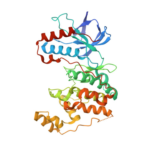
5TCO

Human p38 MAP Kinase in Complex with Dibenzosuberone Compound 1


Experimental Data Snapshot

  • Method: X-RAY DIFFRACTION
  • Resolution: 2.10 Å
  • R-Value Free: 0.270 
  • R-Value Work: 0.214 
  • R-Value Observed: 0.217 

wwPDB Validation   3D Report Full Report


Ligand Structure Quality Assessment 

Created with Raphaël 2.3.0Worse 01 BetterLigand structure goodness of fit to experimental dataBest fitted 79QClick on this verticalbar to view detailsBest fitted BOGClick on this verticalbar to view details

This is version 1.3 of the entry. See complete history


Literature

Optimized Target Residence Time: Type I1/2 Inhibitors for p38 alpha MAP Kinase with Improved Binding Kinetics through Direct Interaction with the R-Spine.

Wentsch, H.K.Walter, N.M.Buhrmann, M.Mayer-Wrangowski, S.Rauh, D.Zaman, G.J.R.Willemsen-Seegers, N.Buijsman, R.C.Henning, M.Dauch, D.Zender, L.Laufer, S.

(2017) Angew Chem Int Ed Engl 56: 5363-5367

  • DOI: https://doi.org/10.1002/anie.201701185
  • Primary Citation of Related Structures:  
    5TBE, 5TCO

  • PubMed Abstract: 

    Skepinone-L was recently reported to be a p38α MAP kinase inhibitor with high potency and excellent selectivity in vitro and in vivo. However, this class of compounds still act as fully ATP-competitive Type I binders which, furthermore, suffer from short residence times at the enzyme. We herein describe a further development with the first Type I1/2 binders for p38α MAP kinase. Type I1/2 inhibitors interfere with the R-spine, inducing a glycine flip and occupying both hydrophobic regions I and II. This design approach leads to prolonged target residence time, binding to both the active and inactive states of the kinase, excellent selectivity, excellent potency on the enzyme level, and low nanomolar activity in a human whole blood assay. This promising binding mode is proven by X-ray crystallography.


  • Organizational Affiliation

    Institute of Pharmaceutical Sciences, Pharmaceutical and Medicinal Chemistry, Eberhard Karls Universität Tübingen, Auf der Morgenstelle 8, 72076, Tübingen, Germany.


Macromolecules
Find similar proteins by:  (by identity cutoff)  |  3D Structure
Entity ID: 1
MoleculeChains Sequence LengthOrganismDetailsImage
Mitogen-activated protein kinase 14360Homo sapiensMutation(s): 0 
Gene Names: MAPK14CSBPCSBP1CSBP2CSPB1MXI2SAPK2A
EC: 2.7.11.24
UniProt & NIH Common Fund Data Resources
Find proteins for Q16539 (Homo sapiens)
Explore Q16539 
Go to UniProtKB:  Q16539
PHAROS:  Q16539
GTEx:  ENSG00000112062 
Entity Groups  
Sequence Clusters30% Identity50% Identity70% Identity90% Identity95% Identity100% Identity
UniProt GroupQ16539
Sequence Annotations
Expand
  • Reference Sequence
Small Molecules
Ligands 2 Unique
IDChains Name / Formula / InChI Key2D Diagram3D Interactions
79Q
Query on 79Q

Download Ideal Coordinates CCD File 
B [auth A]3-[(3-benzamido-4-fluoranyl-phenyl)amino]-~{N}-(2-morpholin-4-ylethyl)-11-oxidanylidene-5,6-dihydrodibenzo[1,2-~{d}:1',2'-~{f}][7]annulene-9-carboxamide
C35 H33 F N4 O4
JWZSSEWMVYKYKW-UHFFFAOYSA-N
BOG
Query on BOG

Download Ideal Coordinates CCD File 
C [auth A],
D [auth A]
octyl beta-D-glucopyranoside
C14 H28 O6
HEGSGKPQLMEBJL-RKQHYHRCSA-N
Binding Affinity Annotations 
IDSourceBinding Affinity
79Q BindingDB:  5TCO Kd: min: 3.1, max: 3.9 (nM) from 2 assay(s)
IC50: min: 0.2, max: 38 (nM) from 5 assay(s)
Experimental Data & Validation

Experimental Data

  • Method: X-RAY DIFFRACTION
  • Resolution: 2.10 Å
  • R-Value Free: 0.270 
  • R-Value Work: 0.214 
  • R-Value Observed: 0.217 
  • Space Group: P 21 21 21
Unit Cell:
Length ( Å )Angle ( ˚ )
a = 66.07α = 90
b = 69.75β = 90
c = 74.73γ = 90
Software Package:
Software NamePurpose
REFMACrefinement
XDSdata reduction
XSCALEdata scaling
PHASERphasing

Structure Validation

View Full Validation Report


Ligand Structure Quality Assessment 

Created with Raphaël 2.3.0Worse 01 BetterLigand structure goodness of fit to experimental dataBest fitted 79QClick on this verticalbar to view detailsBest fitted BOGClick on this verticalbar to view details

Entry History 

Deposition Data

Revision History  (Full details and data files)

  • Version 1.0: 2017-04-19
    Type: Initial release
  • Version 1.1: 2017-05-03
    Changes: Database references
  • Version 1.2: 2020-07-29
    Type: Remediation
    Reason: Carbohydrate remediation
    Changes: Data collection, Derived calculations, Structure summary
  • Version 1.3: 2024-05-08
    Changes: Data collection, Database references, Structure summary