5UFX | pdb_00005ufx

Estrogen Receptor Alpha Ligand Binding Domain in Complex with OP1074


Experimental Data Snapshot

  • Method: X-RAY DIFFRACTION
  • Resolution: 1.55 Å
  • R-Value Free: 
    0.230 (Depositor), 0.230 (DCC) 
  • R-Value Work: 
    0.195 (Depositor), 0.200 (DCC) 
  • R-Value Observed: 
    0.197 (Depositor) 

Starting Model: experimental
View more details

wwPDB Validation   3D Report Full Report


Ligand Structure Quality Assessment 

Created with Raphaël 2.3.0Worse 01 BetterLigand structure goodness of fit to experimental dataBest fitted 86YClick on this verticalbar to view details

This is version 1.2 of the entry. See complete history


Literature

Specific stereochemistry of OP-1074 disrupts estrogen receptor alpha helix 12 and confers pure antiestrogenic activity.

Fanning, S.W.Hodges-Gallagher, L.Myles, D.C.Sun, R.Fowler, C.E.Plant, I.N.Green, B.D.Harmon, C.L.Greene, G.L.Kushner, P.J.

(2018) Nat Commun 9: 2368-2368

  • DOI: https://doi.org/10.1038/s41467-018-04413-3
  • Primary Citation of Related Structures:  
    5UFW, 5UFX, 6C42

  • PubMed Abstract: 

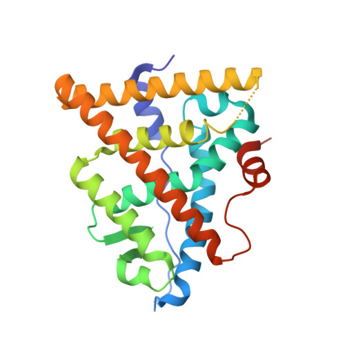
    Complex tissue-specific and cell-specific signaling by the estrogen receptor (ER) frequently leads to the development of resistance to endocrine therapy for breast cancer. Pure ER antagonists, which completely lack tissue-specific agonist activity, hold promise for preventing and treating endocrine resistance, however an absence of structural information hinders the development of novel candidates. Here we synthesize a small panel of benzopyrans with variable side chains to identify pure antiestrogens in a uterotrophic assay. We identify OP-1074 as a pure antiestrogen and a selective ER degrader (PA-SERD) that is efficacious in shrinking tumors in a tamoxifen-resistant xenograft model. Biochemical and crystal structure analyses reveal a structure activity relationship implicating the importance of a stereospecific methyl on the pyrrolidine side chain of OP-1074, particularly on helix 12.


  • Organizational Affiliation

    Ben May Department for Cancer Research, University of Chicago, Chicago, IL, 60637, USA. sfanning@uchicago.edu.


Macromolecules
Find similar proteins by:  (by identity cutoff)  |  3D Structure
Entity ID: 1
MoleculeChains Sequence LengthOrganismDetailsImage
Estrogen receptor
A, B
249Homo sapiensMutation(s): 4 
Gene Names: ESR1ESRNR3A1
UniProt & NIH Common Fund Data Resources
Find proteins for P03372 (Homo sapiens)
Explore P03372 
Go to UniProtKB:  P03372
PHAROS:  P03372
GTEx:  ENSG00000091831 
Entity Groups  
Sequence Clusters30% Identity50% Identity70% Identity90% Identity95% Identity100% Identity
UniProt GroupP03372
Sequence Annotations
Expand
  • Reference Sequence
Small Molecules
Ligands 1 Unique
IDChains Name / Formula / InChI Key2D Diagram3D Interactions
86Y
Query on 86Y

Download Ideal Coordinates CCD File 
C [auth A],
D [auth B]
(2S)-3-(4-hydroxyphenyl)-4-methyl-2-(4-{2-[(3R)-3-methylpyrrolidin-1-yl]ethoxy}phenyl)-2H-1-benzopyran-7-ol
C29 H31 N O4
KDVXAPCZVZMPMU-XBBWARJSSA-N
Experimental Data & Validation

Experimental Data

  • Method: X-RAY DIFFRACTION
  • Resolution: 1.55 Å
  • R-Value Free:  0.230 (Depositor), 0.230 (DCC) 
  • R-Value Work:  0.195 (Depositor), 0.200 (DCC) 
  • R-Value Observed: 0.197 (Depositor) 
Space Group: C 1 2 1
Unit Cell:
Length ( Å )Angle ( ˚ )
a = 102.722α = 90
b = 58.015β = 102.89
c = 87.834γ = 90
Software Package:
Software NamePurpose
PHENIXrefinement
PHENIXdata reduction
PHENIXdata scaling
PHENIXphasing

Structure Validation

View Full Validation Report


Ligand Structure Quality Assessment 

Created with Raphaël 2.3.0Worse 01 BetterLigand structure goodness of fit to experimental dataBest fitted 86YClick on this verticalbar to view details

Entry History 

Deposition Data

Revision History  (Full details and data files)

  • Version 1.0: 2018-01-10
    Type: Initial release
  • Version 1.1: 2018-06-27
    Changes: Data collection, Database references
  • Version 1.2: 2023-10-04
    Changes: Advisory, Data collection, Database references, Refinement description