5UGR | pdb_00005ugr

Malyl-CoA lyase from Methylobacterium extorquens


Experimental Data Snapshot

  • Method: X-RAY DIFFRACTION
  • Resolution: 1.56 Å
  • R-Value Free: 
    0.134 (Depositor), 0.135 (DCC) 
  • R-Value Work: 
    0.102 (Depositor), 0.102 (DCC) 
  • R-Value Observed: 
    0.104 (Depositor) 

Starting Model: experimental
View more details

wwPDB Validation 3D Report Full Report

Validation slider image for 5UGR

This is version 1.3 of the entry. See complete history

Literature

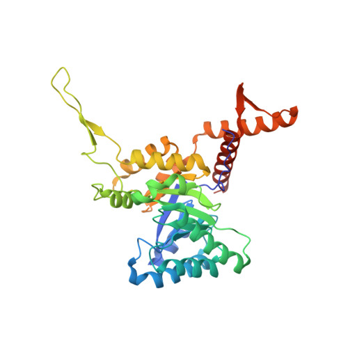
Structure of Methylobacterium extorquens malyl-CoA lyase: CoA-substrate binding correlates with domain shift.

Gonzalez, J.M.Marti-Arbona, R.Chen, J.C.Unkefer, C.J.

(2017) Acta Crystallogr F Struct Biol Commun 73: 79-85

  • DOI: https://doi.org/10.1107/S2053230X17001029
  • Primary Citation Related Structures: 
    5UGR

  • PubMed Abstract: 

    Malyl-CoA lyase (MCL) is an Mg 2+ -dependent enzyme that catalyzes the reversible cleavage of (2S)-4-malyl-CoA to yield acetyl-CoA and glyoxylate. MCL enzymes, which are found in a variety of bacteria, are members of the citrate lyase-like family and are involved in the assimilation of one- and two-carbon compounds. Here, the 1.56 Å resolution X-ray crystal structure of MCL from Methylobacterium extorquens AM1 with bound Mg 2+ is presented. Structural alignment with the closely related Rhodobacter sphaeroides malyl-CoA lyase complexed with Mg 2+ , oxalate and CoA allows a detailed analysis of the domain motion of the enzyme caused by substrate binding. Alignment of the structures shows that a simple hinge motion centered on the conserved residues Phe268 and Thr269 moves the C-terminal domain by about 30° relative to the rest of the molecule. This domain motion positions a conserved aspartate residue located in the C-terminal domain in the active site of the adjacent monomer, which may serve as a general acid/base in the catalytic mechanism.


  • Organizational Affiliation
    • Bioscience Division, Los Alamos National Laboratory, Los Alamos, NM 87545, USA.

Macromolecule Content 

  • Total Structure Weight: 37.03 kDa 
  • Atom Count: 2,963 
  • Modeled Residue Count: 323 
  • Deposited Residue Count: 333 
  • Unique protein chains: 1

Macromolecules

Find similar proteins by:|  3D Structure
Entity ID: 1
MoleculeChains  Sequence LengthOrganismDetailsImage
Malyl-CoA lyase/beta-methylmalyl-CoA lyase333Methylorubrum extorquens AM1Mutation(s): 0 
Gene Names: mclMexAM1_META1p1733
EC: 4.1.3.24
UniProt
Find proteins for C5B113 (Methylorubrum extorquens (strain ATCC 14718 / DSM 1338 / JCM 2805 / NCIMB 9133 / AM1))
Explore C5B113 
Go to UniProtKB:  C5B113
Entity Groups
Sequence Clusters30% Identity50% Identity70% Identity90% Identity95% Identity100% Identity
UniProt GroupC5B113
Sequence Annotations
Expand
Reference Sequence

Experimental Data & Validation

Experimental Data

  • Method: X-RAY DIFFRACTION
  • Resolution: 1.56 Å
  • R-Value Free:  0.134 (Depositor), 0.135 (DCC) 
  • R-Value Work:  0.102 (Depositor), 0.102 (DCC) 
  • R-Value Observed: 0.104 (Depositor) 
Space Group: P 6 2 2
Unit Cell:
Length ( Å )Angle ( ˚ )
a = 126.524α = 90
b = 126.524β = 90
c = 103.207γ = 120
Software Package:
Software NamePurpose
REFMACrefinement
XDSdata reduction
Aimlessdata scaling
PHASERphasing

Structure Validation

View Full Validation Report


Entry History 

Deposition Data

  • Released Date: 2017-02-08 
  • Deposition Author(s): Gonzalez, J.M.
  • This entry supersedes: 4ROQ

Revision History  (Full details and data files)

  • Version 1.0: 2017-02-08
    Type: Initial release
  • Version 1.1: 2017-02-22
    Changes: Database references
  • Version 1.2: 2017-08-23
    Changes: Advisory, Data collection
  • Version 1.3: 2023-10-04
    Changes: Data collection, Database references, Refinement description