5UK8

The co-structure of (R)-4-(6-(1-(cyclopropylsulfonyl)cyclopropyl)-2-(1H-indol-4-yl)pyrimidin-4-yl)-3-methylmorpholine and a rationally designed PI3K-alpha mutant that mimics ATR


Experimental Data Snapshot

  • Method: X-RAY DIFFRACTION
  • Resolution: 2.50 Å
  • R-Value Free: 0.240 
  • R-Value Work: 0.198 
  • R-Value Observed: 0.200 

wwPDB Validation   3D Report Full Report


Ligand Structure Quality Assessment 


This is version 1.2 of the entry. See complete history


Literature

Rationally Designed PI3K alpha Mutants to Mimic ATR and Their Use to Understand Binding Specificity of ATR Inhibitors.

Lu, Y.Knapp, M.Crawford, K.Warne, R.Elling, R.Yan, K.Doyle, M.Pardee, G.Zhang, L.Ma, S.Mamo, M.Ornelas, E.Pan, Y.Bussiere, D.Jansen, J.Zaror, I.Lai, A.Barsanti, P.Sim, J.

(2017) J Mol Biol 429: 1684-1704

  • DOI: https://doi.org/10.1016/j.jmb.2017.04.006
  • Primary Citation of Related Structures:  
    5UK8, 5UKJ, 5UL1

  • PubMed Abstract: 

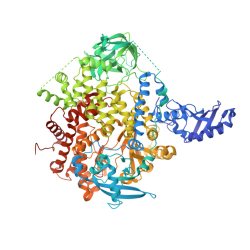
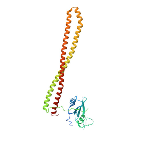
    ATR, a protein kinase in the PIKK family, plays a critical role in the cell DNA-damage response and is an attractive anticancer drug target. Several potent and selective inhibitors of ATR have been reported showing significant antitumor efficacy, with most advanced ones entering clinical trials. However, due to the absence of an experimental ATR structure, the determinants contributing to ATR inhibitors' potency and specificity are not well understood. Here we present the mutations in the ATP-binding site of PI3Kα to progressively transform the pocket to mimic that of ATR. The generated PI3Kα mutants exhibit significantly improved affinity for selective ATR inhibitors in multiple chemical classes. Furthermore, we obtained the X-ray structures of the PI3Kα mutants in complex with the ATR inhibitors. The crystal structures together with the analysis on the inhibitor affinity profile elucidate the roles of individual amino acid residues in the binding of ATR inhibitors, offering key insights for the binding mechanism and revealing the structure features important for the specificity of ATR inhibitors. The ability to obtain structural and binding data for these PI3Kα mutants, together with their ATR-like inhibitor binding profiles, makes these chimeric PI3Kα proteins valuable model systems for structure-based inhibitor design.


  • Organizational Affiliation

    Global Discovery Chemistry, Novartis Institutes for Biomedical Research, Emeryville, CA 94608, USA. Electronic address: yipin.lu@novartis.com.


Macromolecules
Find similar proteins by:  (by identity cutoff)  |  3D Structure
Entity ID: 1
MoleculeChains Sequence LengthOrganismDetailsImage
Phosphatidylinositol 4,5-bisphosphate 3-kinase catalytic subunit alpha isoform1,074Homo sapiensMutation(s): 8 
Gene Names: PIK3CA
EC: 2.7.1.153 (PDB Primary Data), 2.7.11.1 (PDB Primary Data)
UniProt & NIH Common Fund Data Resources
Find proteins for P42336 (Homo sapiens)
Explore P42336 
Go to UniProtKB:  P42336
PHAROS:  P42336
GTEx:  ENSG00000121879 
Entity Groups  
Sequence Clusters30% Identity50% Identity70% Identity90% Identity95% Identity100% Identity
UniProt GroupP42336
Sequence Annotations
Expand
  • Reference Sequence
Find similar proteins by:  (by identity cutoff)  |  3D Structure
Entity ID: 2
MoleculeChains Sequence LengthOrganismDetailsImage
Phosphatidylinositol 3-kinase regulatory subunit alpha293Homo sapiensMutation(s): 1 
Gene Names: PIK3R1GRB1
UniProt & NIH Common Fund Data Resources
Find proteins for P27986 (Homo sapiens)
Explore P27986 
Go to UniProtKB:  P27986
PHAROS:  P27986
GTEx:  ENSG00000145675 
Entity Groups  
Sequence Clusters30% Identity50% Identity70% Identity90% Identity95% Identity100% Identity
UniProt GroupP27986
Sequence Annotations
Expand
  • Reference Sequence
Small Molecules
Ligands 1 Unique
IDChains Name / Formula / InChI Key2D Diagram3D Interactions
8DV
Query on 8DV

Download Ideal Coordinates CCD File 
C [auth A](R)-4-(6-(1-(cyclopropylsulfonyl)cyclopropyl)-2-(1H-indol-4-yl)pyrimidin-4-yl)-3-methylmorpholine
C23 H26 N4 O3 S
NTJNSGIJAQGTSK-OAHLLOKOSA-N
Binding Affinity Annotations 
IDSourceBinding Affinity
8DV Binding MOAD:  5UK8 Kd: 400 (nM) from 1 assay(s)
Experimental Data & Validation

Experimental Data

  • Method: X-RAY DIFFRACTION
  • Resolution: 2.50 Å
  • R-Value Free: 0.240 
  • R-Value Work: 0.198 
  • R-Value Observed: 0.200 
  • Space Group: P 21 21 21
Unit Cell:
Length ( Å )Angle ( ˚ )
a = 104.028α = 90
b = 107.097β = 90
c = 134.706γ = 90
Software Package:
Software NamePurpose
REFMACrefinement
XDSdata reduction
Aimlessdata scaling
PHASERphasing
Cootmodel building
BUSTERrefinement

Structure Validation

View Full Validation Report


Ligand Structure Quality Assessment 


Entry History 

Deposition Data

Revision History  (Full details and data files)

  • Version 1.0: 2017-06-14
    Type: Initial release
  • Version 1.1: 2024-03-06
    Changes: Data collection, Database references
  • Version 1.2: 2024-04-03
    Changes: Refinement description