5V0N | pdb_00005v0n

BACE1 in complex with inhibitor 5g


Experimental Data Snapshot

  • Method: X-RAY DIFFRACTION
  • Resolution: 2.15 Å
  • R-Value Free: 
    0.202 (Depositor), 0.210 (DCC) 
  • R-Value Work: 
    0.161 (Depositor), 0.169 (DCC) 
  • R-Value Observed: 
    0.162 (Depositor) 

Starting Model: experimental
View more details

wwPDB Validation 3D Report Full Report

Validation slider image for 5V0N

Ligand Structure Quality Assessment 


This is version 1.3 of the entry. See complete history

Literature

Design, synthesis, and X-ray structural studies of BACE-1 inhibitors containing substituted 2-oxopiperazines as P1'-P2' ligands.

Ghosh, A.K.Brindisi, M.Yen, Y.C.Cardenas, E.L.Ella-Menye, J.R.Kumaragurubaran, N.Huang, X.Tang, J.Mesecar, A.D.

(2017) Bioorg Med Chem Lett 27: 2432-2438

  • DOI: https://doi.org/10.1016/j.bmcl.2017.04.011
  • Primary Citation Related Structures: 
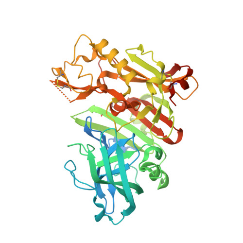
    5V0N

  • PubMed Abstract: 

    We report the design and synthesis of a series of BACE1 inhibitors incorporating mono- and bicyclic 6-substituted 2-oxopiperazines as novel P1' and P2' ligands and isophthalamide derivative as P2-P3 ligands. Among mono-substituted 2-oxopiperazines, inhibitor 5a with N-benzyl-2-oxopiperazine and isophthalamide showed potent BACE1 inhibitory activity (K i =2nM). Inhibitor 5g, with N-benzyl-2-oxopiperazine and substituted indole-derived P2-ligand showed a reduction in potency. The X-ray crystal structure of 5g-bound BACE1 was determined and used to design a set of disubstituted 2-oxopiperazines and bicyclic derivatives that were subsequently investigated. Inhibitor 6j with an oxazolidinone derivative showed a BACE1 inhibitory activity of 23nM and cellular EC 50 of 80nM.


  • Organizational Affiliation
    • Department of Chemistry, Purdue University, West Lafayette, IN 47907, United States; Department of Medicinal Chemistry, Purdue University, West Lafayette, IN 47907, United States. Electronic address: akghosh@purdue.edu.

Macromolecule Content 

  • Total Structure Weight: 149.76 kDa 
  • Atom Count: 9,715 
  • Modeled Residue Count: 1,121 
  • Deposited Residue Count: 1,323 
  • Unique protein chains: 1

Macromolecules

Find similar proteins by:|  3D Structure
Entity ID: 1
MoleculeChains  Sequence LengthOrganismDetailsImage
Beta-secretase 1
A, B, C
441Homo sapiensMutation(s): 0 
Gene Names: BACE1BACEKIAA1149
EC: 3.4.23.46
UniProt & NIH Common Fund Data Resources
Find proteins for P56817 (Homo sapiens)
Explore P56817 
Go to UniProtKB:  P56817
PHAROS:  P56817
GTEx:  ENSG00000186318 
Entity Groups
Sequence Clusters30% Identity50% Identity70% Identity90% Identity95% Identity100% Identity
UniProt GroupP56817
Sequence Annotations
Expand
Reference Sequence

Small Molecules

Ligands 4 Unique
IDChains Name / Formula / InChI Key2D Diagram3D Interactions
8W4

Query on 8W4


Download:Ideal Coordinates CCD File
G [auth A],
N [auth B],
T [auth C]
N-{(1S,2S)-1-[(2S)-4-benzyl-3-oxopiperazin-2-yl]-1-hydroxy-3-phenylpropan-2-yl}-7-ethyl-1,3,3-trimethyl-2,2-dioxo-1,2,3,4-tetrahydro-2lambda~6~-[1,2,5]thiadiazepino[3,4,5-hi]indole-9-carboxamide
C36 H43 N5 O5 S
WBWSVNAKBNLSOJ-WXWREJFYSA-N
SO4

Query on SO4


Download:Ideal Coordinates CCD File
D [auth A]
E [auth A]
F [auth A]
I [auth B]
J [auth B]
D [auth A],
E [auth A],
F [auth A],
I [auth B],
J [auth B],
K [auth B],
L [auth B],
P [auth C],
Q [auth C],
R [auth C],
S [auth C]
SULFATE ION
O4 S
QAOWNCQODCNURD-UHFFFAOYSA-L
GOL

Query on GOL


Download:Ideal Coordinates CCD File
M [auth B]GLYCEROL
C3 H8 O3
PEDCQBHIVMGVHV-UHFFFAOYSA-N
URE

Query on URE


Download:Ideal Coordinates CCD File
H [auth A],
O [auth B]
UREA
C H4 N2 O
XSQUKJJJFZCRTK-UHFFFAOYSA-N
Binding Affinity Annotations 
IDSourceBinding Affinity
8W4 BindingDB:  5V0N Ki: 67 (nM) from 1 assay(s)
EC50: 19 (nM) from 1 assay(s)

Experimental Data & Validation

Experimental Data

  • Method: X-RAY DIFFRACTION
  • Resolution: 2.15 Å
  • R-Value Free:  0.202 (Depositor), 0.210 (DCC) 
  • R-Value Work:  0.161 (Depositor), 0.169 (DCC) 
  • R-Value Observed: 0.162 (Depositor) 
Space Group: P 1 21 1
Unit Cell:
Length ( Å )Angle ( ˚ )
a = 82.04α = 90
b = 103.545β = 104.59
c = 99.765γ = 90
Software Package:
Software NamePurpose
PHENIXrefinement
MOSFLMdata reduction
SCALAdata scaling
PHASERphasing

Structure Validation

View Full Validation Report


Ligand Structure Quality Assessment 


Entry History 

Deposition Data

Revision History  (Full details and data files)

  • Version 1.0: 2017-05-03
    Type: Initial release
  • Version 1.1: 2017-05-24
    Changes: Database references
  • Version 1.2: 2023-10-04
    Changes: Data collection, Database references, Refinement description
  • Version 1.3: 2024-11-20
    Changes: Structure summary