


Crystal structure of an alpha,alpha-trehalose-phosphate synthase (UDP-forming) from Burkholderia xenovorans in complex with glucose-6-phosphate
Mayclin, S.J., Dranow, D.M., Lorimer, D., Edwards, T.E.To be published.
Experimental Data Snapshot
Starting Model: experimental
View more details
Macromolecule Content
Entity ID: 1 | |||||
|---|---|---|---|---|---|
| Molecule | Chains | Sequence Length | Organism | Details | Image |
| Alpha,alpha-trehalose-phosphate synthase (UDP-forming) | 494 | Paraburkholderia xenovorans LB400 | Mutation(s): 0 Gene Names: Bxe_A1242 EC: 2.4.1.15 | 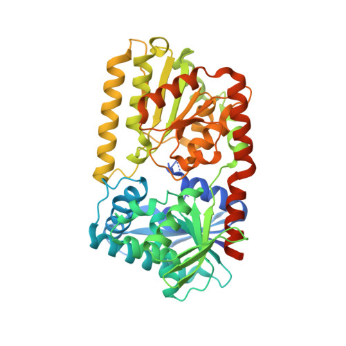 | |
UniProt | |||||
Entity Groups | |||||
| Sequence Clusters | 30% Identity50% Identity70% Identity90% Identity95% Identity100% Identity | ||||
| UniProt Group | Q13W28 | ||||
Sequence AnnotationsExpand | |||||
Reference Sequence | |||||
| Ligands 7 Unique | |||||
|---|---|---|---|---|---|
| ID | Chains | Name / Formula / InChI Key | 2D Diagram | 3D Interactions | |
| UDP Download:Ideal Coordinates CCD File | CC [auth H] EB [auth E] I [auth A] MA [auth C] PB [auth F] | URIDINE-5'-DIPHOSPHATE C9 H14 N2 O12 P2 XCCTYIAWTASOJW-XVFCMESISA-N |  | ||
| G6P Download:Ideal Coordinates CCD File | DC [auth H] FB [auth E] J [auth A] NA [auth C] QB [auth F] | 6-O-phosphono-alpha-D-glucopyranose C6 H13 O9 P NBSCHQHZLSJFNQ-DVKNGEFBSA-N |  | ||
| TAR Download:Ideal Coordinates CCD File | GB [auth E] | D(-)-TARTARIC ACID C4 H6 O6 FEWJPZIEWOKRBE-LWMBPPNESA-N |  | ||
| OXD Download:Ideal Coordinates CCD File | EA [auth B], JB [auth E], O [auth A], RA [auth C], UB [auth F] | OXALIC ACID C2 H2 O4 MUBZPKHOEPUJKR-UHFFFAOYSA-N |  | ||
| EDO Download:Ideal Coordinates CCD File | AC [auth G] EC [auth H] FC [auth H] HB [auth E] K [auth A] | 1,2-ETHANEDIOL C2 H6 O2 LYCAIKOWRPUZTN-UHFFFAOYSA-N |  | ||
| ACT Download:Ideal Coordinates CCD File | AA [auth B] BA [auth B] BC [auth G] CA [auth B] DA [auth B] | ACETATE ION C2 H3 O2 QTBSBXVTEAMEQO-UHFFFAOYSA-M |  | ||
| FMT Download:Ideal Coordinates CCD File | AB [auth D] BB [auth D] CB [auth D] DB [auth D] FA [auth B] | FORMIC ACID C H2 O2 BDAGIHXWWSANSR-UHFFFAOYSA-N |  | ||
| Length ( Å ) | Angle ( ˚ ) |
|---|---|
| a = 84.92 | α = 91.36 |
| b = 105.95 | β = 90.37 |
| c = 135.71 | γ = 89.97 |
| Software Name | Purpose |
|---|---|
| XSCALE | data scaling |
| Coot | model building |
| PHENIX | refinement |
| PDB_EXTRACT | data extraction |
| XDS | data reduction |
| MOLREP | phasing |


