5V6V | pdb_00005v6v

Crystal structure of small molecule aziridine 3 covalently bound to K-Ras G12C


Experimental Data Snapshot

  • Method: X-RAY DIFFRACTION
  • Resolution: 1.72 Å
  • R-Value Free: 
    0.202 (Depositor), 0.202 (DCC) 
  • R-Value Work: 
    0.168 (Depositor), 0.170 (DCC) 
  • R-Value Observed: 
    0.170 (Depositor) 

wwPDB Validation 3D Report Full Report

Validation slider image for 5V6V

Ligand Structure Quality Assessment 


This is version 1.4 of the entry. See complete history

Literature

Expanding the Scope of Electrophiles Capable of Targeting K-Ras Oncogenes.

McGregor, L.M.Jenkins, M.L.Kerwin, C.Burke, J.E.Shokat, K.M.

(2017) Biochemistry 56: 3178-3183

  • DOI: https://doi.org/10.1021/acs.biochem.7b00271
  • Primary Citation Related Structures: 
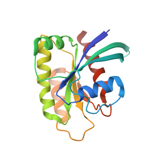
    5V6S, 5V6V

  • PubMed Abstract: 

    There is growing interest in reversible and irreversible covalent inhibitors that target noncatalytic amino acids in target proteins. With a goal of targeting oncogenic K-Ras variants (e.g., G12D) by expanding the types of amino acids that can be targeted by covalent inhibitors, we survey a set of electrophiles for their ability to label carboxylates. We functionalized an optimized ligand for the K-Ras switch II pocket with a set of electrophiles previously reported to react with carboxylates and characterized the ability of these compounds to react with model nucleophiles and oncogenic K-Ras proteins. Here, we report that aziridines and stabilized diazo groups preferentially react with free carboxylates over thiols. Although we did not identify a warhead that potently labels K-Ras G12D, we were able to study the interactions of many electrophiles with K-Ras, as most of the electrophiles rapidly label K-Ras G12C. We characterized the resulting complexes by crystallography, hydrogen/deuterium exchange, and differential scanning fluorimetry. Our results both demonstrate the ability of a noncatalytic cysteine to react with a diverse set of electrophiles and emphasize the importance of proper spatial arrangements between a covalent inhibitor and its intended nucleophile. We hope that these results can expand the range of electrophiles and nucleophiles of use in covalent protein modulation.


  • Organizational Affiliation
    • Howard Hughes Medical Institute and Department of Cellular and Molecular Pharmacology, University of California, San Francisco , San Francisco, California 94158, United States.

Macromolecule Content 

  • Total Structure Weight: 40.86 kDa 
  • Atom Count: 3,086 
  • Modeled Residue Count: 333 
  • Deposited Residue Count: 340 
  • Unique protein chains: 1

Macromolecules

Find similar proteins by:|  3D Structure
Entity ID: 1
MoleculeChains  Sequence LengthOrganismDetailsImage
GTPase KRas
A, B
170Homo sapiensMutation(s): 4 
Gene Names: KRASKRAS2RASK2
EC: 3.6.5.2
UniProt & NIH Common Fund Data Resources
Find proteins for P01116 (Homo sapiens)
Explore P01116 
Go to UniProtKB:  P01116
PHAROS:  P01116
GTEx:  ENSG00000133703 
Entity Groups
Sequence Clusters30% Identity50% Identity70% Identity90% Identity95% Identity100% Identity
UniProt GroupP01116
Sequence Annotations
Expand
Reference Sequence

Small Molecules

Ligands 4 Unique
IDChains Name / Formula / InChI Key2D Diagram3D Interactions
8YA

Query on 8YA


Download:Ideal Coordinates CCD File
F [auth A],
M [auth B]
3-amino-1-{4-[6-chloro-8-fluoro-7-(5-methyl-1H-indazol-4-yl)quinazolin-4-yl]piperazin-1-yl}propan-1-one
C23 H23 Cl F N7 O
VJDYKBMYNVLSKK-UHFFFAOYSA-N
GDP

Query on GDP


Download:Ideal Coordinates CCD File
C [auth A],
H [auth B]
GUANOSINE-5'-DIPHOSPHATE
C10 H15 N5 O11 P2
QGWNDRXFNXRZMB-UUOKFMHZSA-N
GOL

Query on GOL


Download:Ideal Coordinates CCD File
L [auth B]GLYCEROL
C3 H8 O3
PEDCQBHIVMGVHV-UHFFFAOYSA-N
CA

Query on CA


Download:Ideal Coordinates CCD File
D [auth A]
E [auth A]
G [auth B]
I [auth B]
J [auth B]
D [auth A],
E [auth A],
G [auth B],
I [auth B],
J [auth B],
K [auth B]
CALCIUM ION
Ca
BHPQYMZQTOCNFJ-UHFFFAOYSA-N

Experimental Data & Validation

Experimental Data

  • Method: X-RAY DIFFRACTION
  • Resolution: 1.72 Å
  • R-Value Free:  0.202 (Depositor), 0.202 (DCC) 
  • R-Value Work:  0.168 (Depositor), 0.170 (DCC) 
  • R-Value Observed: 0.170 (Depositor) 
Space Group: P 1
Unit Cell:
Length ( Å )Angle ( ˚ )
a = 33.23α = 77.34
b = 39.17β = 81.44
c = 62.34γ = 76.86
Software Package:
Software NamePurpose
PHENIXrefinement
PDB_EXTRACTdata extraction
iMOSFLMdata reduction
SCALAdata scaling
PHASERphasing

Structure Validation

View Full Validation Report


Ligand Structure Quality Assessment 


Entry History 

& Funding Information

Deposition Data


Funding OrganizationLocationGrant Number
National Institutes of Health/National Cancer Institute (NIH/NCI)United States5R01CA190408
Howard Hughes Medical Institute (HHMI)United States--

Revision History  (Full details and data files)

  • Version 1.0: 2017-06-28
    Type: Initial release
  • Version 1.1: 2017-07-05
    Changes: Database references
  • Version 1.2: 2017-09-20
    Changes: Author supporting evidence
  • Version 1.3: 2019-11-20
    Changes: Author supporting evidence
  • Version 1.4: 2024-10-23
    Changes: Data collection, Database references, Derived calculations, Structure summary