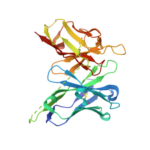
5VF2

scFv 2D10 re-refined as a complex with trehalose replacing the original alpha-1,6-mannobiose


Experimental Data Snapshot

  • Method: X-RAY DIFFRACTION
  • Resolution: 1.55 Å
  • R-Value Free: 0.180 
  • R-Value Work: 0.153 
  • R-Value Observed: 0.156 

Starting Model: experimental
View more details

wwPDB Validation   3D Report Full Report


This is version 2.3 of the entry. See complete history

Re-refinement Note

This entry reflects an alternative modeling of the original data in: 5I4F


Literature

Detect, correct, retract: How to manage incorrect structural models.

Wlodawer, A.Dauter, Z.Porebski, P.J.Minor, W.Stanfield, R.Jaskolski, M.Pozharski, E.Weichenberger, C.X.Rupp, B.

(2018) FEBS J 285: 444-466

  • DOI: https://doi.org/10.1111/febs.14320
  • Primary Citation of Related Structures:  
    5VEH, 5VEP, 5VEQ, 5VER, 5VET, 5VF2, 5VF5, 5VGA

  • PubMed Abstract: 

    The massive technical and computational progress of biomolecular crystallography has generated some adverse side effects. Most crystal structure models, produced by crystallographers or well-trained structural biologists, constitute useful sources of information, but occasional extreme outliers remind us that the process of structure determination is not fail-safe. The occurrence of severe errors or gross misinterpretations raises fundamental questions: Why do such aberrations emerge in the first place? How did they evade the sophisticated validation procedures which often produce clear and dire warnings, and why were severe errors not noticed by the depositors themselves, their supervisors, referees and editors? Once detected, what can be done to either correct, improve or eliminate such models? How do incorrect models affect the underlying claims or biomedical hypotheses they were intended, but failed, to support? What is the long-range effect of the propagation of such errors? And finally, what mechanisms can be envisioned to restore the validity of the scientific record and, if necessary, retract publications that are clearly invalidated by the lack of experimental evidence? We suggest that cognitive bias and flawed epistemology are likely at the root of the problem. By using examples from the published literature and from public repositories such as the Protein Data Bank, we provide case summaries to guide correction or improvement of structural models. When strong claims are unsustainable because of a deficient crystallographic model, removal of such a model and even retraction of the affected publication are necessary to restore the integrity of the scientific record.


  • Organizational Affiliation

    Protein Structure Section, Macromolecular Crystallography Laboratory, National Cancer Institute, Frederick, MD, USA.


Macromolecules
Find similar proteins by:  (by identity cutoff)  |  3D Structure
Entity ID: 1
MoleculeChains Sequence LengthOrganismDetailsImage
scFv 2D10251Mus musculusMutation(s): 0 
Entity Groups  
Sequence Clusters30% Identity50% Identity70% Identity90% Identity95% Identity100% Identity
Sequence Annotations
Expand
  • Reference Sequence
Oligosaccharides

Help

Entity ID: 2
MoleculeChains Length2D Diagram Glycosylation3D Interactions
alpha-D-glucopyranose-(1-1)-alpha-D-glucopyranose
B, C
2N/A
Glycosylation Resources
GlyTouCan:  G92130SN
GlyCosmos:  G92130SN
Small Molecules
Ligands 3 Unique
IDChains Name / Formula / InChI Key2D Diagram3D Interactions
MES
Query on MES

Download Ideal Coordinates CCD File 
D [auth A]2-(N-MORPHOLINO)-ETHANESULFONIC ACID
C6 H13 N O4 S
SXGZJKUKBWWHRA-UHFFFAOYSA-N
MG
Query on MG

Download Ideal Coordinates CCD File 
TA [auth A]MAGNESIUM ION
Mg
JLVVSXFLKOJNIY-UHFFFAOYSA-N
UNX
Query on UNX

Download Ideal Coordinates CCD File 
AA [auth A]
BA [auth A]
CA [auth A]
DA [auth A]
E [auth A]
AA [auth A],
BA [auth A],
CA [auth A],
DA [auth A],
E [auth A],
EA [auth A],
F [auth A],
FA [auth A],
G [auth A],
GA [auth A],
H [auth A],
HA [auth A],
I [auth A],
IA [auth A],
J [auth A],
JA [auth A],
K [auth A],
KA [auth A],
L [auth A],
LA [auth A],
M [auth A],
MA [auth A],
N [auth A],
NA [auth A],
O [auth A],
OA [auth A],
P [auth A],
PA [auth A],
Q [auth A],
QA [auth A],
R [auth A],
RA [auth A],
S [auth A],
SA [auth A],
T [auth A],
U [auth A],
V [auth A],
W [auth A],
X [auth A],
Y [auth A],
Z [auth A]
UNKNOWN ATOM OR ION
X
Biologically Interesting Molecules (External Reference) 1 Unique
Entity ID: 2
IDChains NameType/Class2D Diagram3D Interactions
PRD_900006
Query on PRD_900006
B, C
trehaloseOligosaccharide / Nutrient
Experimental Data & Validation

Experimental Data

  • Method: X-RAY DIFFRACTION
  • Resolution: 1.55 Å
  • R-Value Free: 0.180 
  • R-Value Work: 0.153 
  • R-Value Observed: 0.156 
  • Space Group: P 31 2 1
Unit Cell:
Length ( Å )Angle ( ˚ )
a = 81.093α = 90
b = 81.093β = 90
c = 74.388γ = 120
Software Package:
Software NamePurpose
REFMACrefinement
HKL-3000refinement

Structure Validation

View Full Validation Report


Entry History & Funding Information

Deposition Data


Funding OrganizationLocationGrant Number
National Institutes of Health/National Institute of General Medical Sciences (NIH/NIGMS)United StatesU01HG008424, R01GM117080, R01GM117325
Austrian Science FundAustriaP28395-B26
Polish National Science CentrePoland2013/10/M/NZ1/00251

Revision History  (Full details and data files)

  • Version 1.0: 2017-12-06
    Type: Initial release
  • Version 1.1: 2018-02-14
    Changes: Database references
  • Version 1.2: 2018-03-21
    Changes: Structure summary
  • Version 1.3: 2020-01-01
    Changes: Author supporting evidence
  • Version 2.0: 2020-07-29
    Type: Remediation
    Reason: Carbohydrate remediation
    Changes: Atomic model, Data collection, Derived calculations, Non-polymer description, Structure summary
  • Version 2.1: 2022-04-13
    Changes: Database references, Structure summary
  • Version 2.2: 2023-10-04
    Changes: Data collection, Refinement description
  • Version 2.3: 2024-10-23
    Changes: Structure summary