5VIF

Electrophilic probes for deciphering substrate recognition by O-GlcNAc transferase


Experimental Data Snapshot

  • Method: X-RAY DIFFRACTION
  • Resolution: 2.25 Å
  • R-Value Free: 0.228 
  • R-Value Work: 0.182 
  • R-Value Observed: 0.184 

wwPDB Validation   3D Report Full Report


Ligand Structure Quality Assessment 


This is version 2.1 of the entry. See complete history


Literature

Electrophilic probes for deciphering substrate recognition by O-GlcNAc transferase.

Hu, C.W.Worth, M.Fan, D.Li, B.Li, H.Lu, L.Zhong, X.Lin, Z.Wei, L.Ge, Y.Li, L.Jiang, J.

(2017) Nat Chem Biol 13: 1267-1273

  • DOI: https://doi.org/10.1038/nchembio.2494
  • Primary Citation of Related Structures:  
    5VIE, 5VIF

  • PubMed Abstract: 

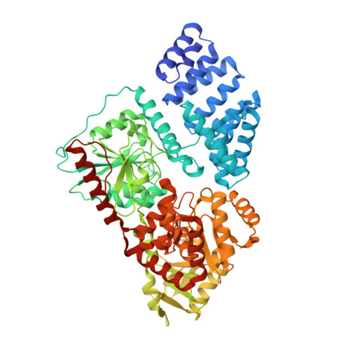
    O-linked β-N-acetylglucosamine (O-GlcNAc) transferase (OGT) is an essential human glycosyltransferase that adds O-GlcNAc modifications to numerous proteins. However, little is known about the mechanism with which OGT recognizes various protein substrates. Here we report on GlcNAc electrophilic probes (GEPs) to expedite the characterization of OGT-substrate recognition. Data from mass spectrometry, X-ray crystallization, and biochemical and radiolabeled kinetic assays support the application of GEPs to rapidly report the impacts of OGT mutations on protein substrate or sugar binding and to discover OGT residues crucial for protein recognition. Interestingly, we found that the same residues on the inner surface of the N-terminal domain contribute to OGT interactions with different protein substrates. By tuning reaction conditions, a GEP enables crosslinking of OGT with acceptor substrates in situ, affording a unique method to discover genuine substrates that weakly or transiently interact with OGT. Hence, GEPs provide new strategies to dissect OGT-substrate binding and recognition.


  • Organizational Affiliation

    Pharmaceutical Sciences Division, School of Pharmacy, University of Wisconsin-Madison, Madison, Wisconsin, USA.


Macromolecules
Find similar proteins by:  (by identity cutoff)  |  3D Structure
Entity ID: 1
MoleculeChains Sequence LengthOrganismDetailsImage
UDP-N-acetylglucosamine--peptide N-acetylglucosaminyltransferase 110 kDa subunit723Homo sapiensMutation(s): 0 
Gene Names: OGT
EC: 2.4.1.255
UniProt & NIH Common Fund Data Resources
Find proteins for O15294 (Homo sapiens)
Explore O15294 
Go to UniProtKB:  O15294
PHAROS:  O15294
GTEx:  ENSG00000147162 
Entity Groups  
Sequence Clusters30% Identity50% Identity70% Identity90% Identity95% Identity100% Identity
UniProt GroupO15294
Sequence Annotations
Expand
  • Reference Sequence

Find similar proteins by:  Sequence   |   3D Structure  

Entity ID: 2
MoleculeChains Sequence LengthOrganismDetailsImage
CKII14Homo sapiensMutation(s): 0 
UniProt & NIH Common Fund Data Resources
Find proteins for P68400 (Homo sapiens)
Explore P68400 
Go to UniProtKB:  P68400
PHAROS:  P68400
GTEx:  ENSG00000101266 
Entity Groups  
Sequence Clusters30% Identity50% Identity70% Identity90% Identity95% Identity100% Identity
UniProt GroupP68400
Sequence Annotations
Expand
  • Reference Sequence
Small Molecules
Ligands 2 Unique
IDChains Name / Formula / InChI Key2D Diagram3D Interactions
UDP
Query on UDP

Download Ideal Coordinates CCD File 
C [auth A]URIDINE-5'-DIPHOSPHATE
C9 H14 N2 O12 P2
XCCTYIAWTASOJW-XVFCMESISA-N
9C1
Query on 9C1

Download Ideal Coordinates CCD File 
D [auth B]2-{[(2E)-4-chlorobut-2-enoyl]amino}-2-deoxy-beta-D-glucopyranose
C10 H16 Cl N O6
LKECTNVLFDGIBK-KKRTVAFTSA-N
Binding Affinity Annotations 
IDSourceBinding Affinity
UDP BindingDB:  5VIF IC50: 1800 (nM) from 1 assay(s)
Experimental Data & Validation

Experimental Data

  • Method: X-RAY DIFFRACTION
  • Resolution: 2.25 Å
  • R-Value Free: 0.228 
  • R-Value Work: 0.182 
  • R-Value Observed: 0.184 
  • Space Group: F 2 2 2
Unit Cell:
Length ( Å )Angle ( ˚ )
a = 139.084α = 90
b = 152.733β = 90
c = 199.398γ = 90
Software Package:
Software NamePurpose
REFMACrefinement
HKL-2000data reduction
HKL-2000data scaling
PHASERphasing

Structure Validation

View Full Validation Report


Ligand Structure Quality Assessment 


Entry History & Funding Information

Deposition Data


Funding OrganizationLocationGrant Number
University of Wisconsin-MadisonUnited States--

Revision History  (Full details and data files)

  • Version 1.0: 2017-10-18
    Type: Initial release
  • Version 1.1: 2017-11-08
    Changes: Database references
  • Version 1.2: 2017-12-06
    Changes: Database references
  • Version 2.0: 2020-07-29
    Type: Remediation
    Reason: Carbohydrate remediation
    Changes: Advisory, Atomic model, Data collection, Derived calculations
  • Version 2.1: 2023-10-04
    Changes: Data collection, Database references, Derived calculations, Refinement description, Structure summary