5VIQ | pdb_00005viq

Crystal structure of monomeric near-infrared fluorescent protein miRFP709


Experimental Data Snapshot

  • Method: X-RAY DIFFRACTION
  • Resolution: 1.34 Å
  • R-Value Free: 
    0.188 (Depositor), 0.188 (DCC) 
  • R-Value Work: 
    0.136 (Depositor), 0.136 (DCC) 
  • R-Value Observed: 
    0.137 (Depositor) 

Starting Model: experimental
View more details

wwPDB Validation 3D Report Full Report

Validation slider image for 5VIQ

Ligand Structure Quality Assessment 


This is version 1.3 of the entry. See complete history

Literature

Designing brighter near-infrared fluorescent proteins: insights from structural and biochemical studies.

Baloban, M.Shcherbakova, D.M.Pletnev, S.Pletnev, V.Z.Lagarias, J.C.Verkhusha, V.V.

(2017) Chem Sci 8: 4546-4557

  • DOI: https://doi.org/10.1039/c7sc00855d
  • Primary Citation Related Structures: 
    5VIK, 5VIQ, 5VIV

  • PubMed Abstract: 

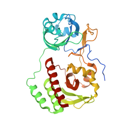
    Brighter near-infrared (NIR) fluorescent proteins (FPs) are required for multicolor microscopy and deep-tissue imaging. Here, we present structural and biochemical analyses of three monomeric, spectrally distinct phytochrome-based NIR FPs, termed miRFPs. The miRFPs are closely related and differ by only a few amino acids, which define their molecular brightness, brightness in mammalian cells, and spectral properties. We have identified the residues responsible for the spectral red-shift, revealed a new chromophore bound simultaneously to two cysteine residues in the PAS and GAF domains in blue-shifted NIR FPs, and uncovered the importance of amino acid residues in the N-terminus of NIR FPs for their molecular and cellular brightness. The novel chromophore covalently links the N-terminus of NIR FPs with their C-terminal GAF domain, forming a topologically closed knot in the structure, and also contributes to the increased brightness. Based on our studies, we suggest a strategy to develop spectrally distinct NIR FPs with enhanced brightness.


  • Organizational Affiliation
    • Department of Anatomy and Structural Biology and Gruss-Lipper Biophotonics Center , Albert Einstein College of Medicine , Bronx , New York 10461 , USA . Email: vladislav.verkhusha@einstein.yu.edu.

Macromolecule Content 

  • Total Structure Weight: 35.19 kDa 
  • Atom Count: 2,768 
  • Modeled Residue Count: 296 
  • Deposited Residue Count: 315 
  • Unique protein chains: 1

Macromolecules

Find similar proteins by:|  3D Structure
Entity ID: 1
MoleculeChains  Sequence LengthOrganismDetailsImage
monomeric near-infrared fluorescent protein miRFP709315Rhodopseudomonas palustrisMutation(s): 0 

Small Molecules

Ligands 1 Unique
IDChains Name / Formula / InChI Key2D Diagram3D Interactions
BLA

Query on BLA


Download:Ideal Coordinates CCD File
B [auth A]BILIVERDINE IX ALPHA
C33 H34 N4 O6
GWZYPXHJIZCRAJ-SRVCBVSDSA-N

Experimental Data & Validation

Experimental Data

  • Method: X-RAY DIFFRACTION
  • Resolution: 1.34 Å
  • R-Value Free:  0.188 (Depositor), 0.188 (DCC) 
  • R-Value Work:  0.136 (Depositor), 0.136 (DCC) 
  • R-Value Observed: 0.137 (Depositor) 
Space Group: P 1 21 1
Unit Cell:
Length ( Å )Angle ( ˚ )
a = 36.266α = 90
b = 52.86β = 92.7
c = 64.625γ = 90
Software Package:
Software NamePurpose
SCALEPACKdata scaling
REFMACrefinement
PDB_EXTRACTdata extraction
DENZOdata reduction
MOLREPphasing

Structure Validation

View Full Validation Report


Ligand Structure Quality Assessment 


Entry History 

Deposition Data

  • Released Date: 2017-06-07 
  • Deposition Author(s): Pletnev, S.

Revision History  (Full details and data files)

  • Version 1.0: 2017-06-07
    Type: Initial release
  • Version 1.1: 2017-10-04
    Changes: Database references
  • Version 1.2: 2023-10-04
    Changes: Data collection, Database references, Refinement description
  • Version 1.3: 2024-10-16
    Changes: Structure summary