5WHA | pdb_00005wha

KRas G12V, bound to GDP and miniprotein 225-11


Experimental Data Snapshot

  • Method: X-RAY DIFFRACTION
  • Resolution: 2.04 Å
  • R-Value Free: 
    0.277 (Depositor), 0.261 (DCC) 
  • R-Value Work: 
    0.238 (Depositor), 0.236 (DCC) 
  • R-Value Observed: 
    0.240 (Depositor) 

Starting Model: experimental
View more details

wwPDB Validation 3D Report Full Report

Validation slider image for 5WHA

Ligand Structure Quality Assessment 


This is version 1.4 of the entry. See complete history

Literature

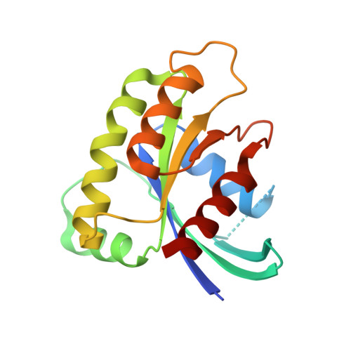
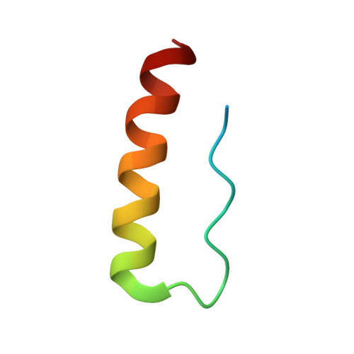
Exceptionally high-affinity Ras binders that remodel its effector domain.

McGee, J.H.Shim, S.Y.Lee, S.J.Swanson, P.K.Jiang, S.Y.Durney, M.A.Verdine, G.L.

(2018) J Biological Chem 293: 3265-3280

  • DOI: https://doi.org/10.1074/jbc.M117.816348
  • Primary Citation Related Structures: 
    5WHA, 5WHB, 5WHD, 5WHE, 5WLB, 5WPL, 5WPM

  • PubMed Abstract: 

    The Ras proteins are aberrantly activated in a wide range of human cancers, often endowing tumors with aggressive properties and resistance to therapy. Decades of effort to develop direct Ras inhibitors for clinical use have thus far failed, largely because of a lack of adequate small-molecule-binding pockets on the Ras surface. Here, we report the discovery of Ras-binding miniproteins from a naïve library and their evolution to afford versions with midpicomolar affinity to Ras. A series of biochemical experiments indicated that these miniproteins bind to the Ras effector domain as dimers, and high-resolution crystal structures revealed that these miniprotein dimers bind Ras in an unprecedented mode in which the Ras effector domain is remodeled to expose an extended pocket that connects two isolated pockets previously found to engage small-molecule ligands. We also report a Ras point mutant that stabilizes the protein in the open conformation trapped by these miniproteins. These findings provide new tools for studying Ras structure and function and present opportunities for the development of both miniprotein and small-molecule inhibitors that directly target the Ras proteins.


  • Organizational Affiliation
    • From the Departments of Molecular and Cellular Biology.

Macromolecule Content 

  • Total Structure Weight: 110.79 kDa 
  • Atom Count: 7,278 
  • Modeled Residue Count: 858 
  • Deposited Residue Count: 960 
  • Unique protein chains: 2

Macromolecules

Find similar proteins by:|  3D Structure
Entity ID: 1
MoleculeChains  Sequence LengthOrganismDetailsImage
GTPase KRas
A, D, G, J
170Homo sapiensMutation(s): 1 
Gene Names: KRASKRAS2RASK2
EC: 3.6.5.2
UniProt & NIH Common Fund Data Resources
Find proteins for P01116 (Homo sapiens)
Explore P01116 
Go to UniProtKB:  P01116
PHAROS:  P01116
GTEx:  ENSG00000133703 
Entity Groups
Sequence Clusters30% Identity50% Identity70% Identity90% Identity95% Identity100% Identity
UniProt GroupP01116
Sequence Annotations
Expand
Reference Sequence
Find similar proteins by:|  3D Structure
Entity ID: 2
MoleculeChains  Sequence LengthOrganismDetailsImage
miniprotein 225-11
B, C, E, F, H
B, C, E, F, H, I, K, L
35synthetic constructMutation(s): 0 

Small Molecules

Ligands 3 Unique
IDChains Name / Formula / InChI Key2D Diagram3D Interactions
GDP

Query on GDP


Download:Ideal Coordinates CCD File
BA [auth J],
N [auth A],
R [auth D],
V [auth G]
GUANOSINE-5'-DIPHOSPHATE
C10 H15 N5 O11 P2
QGWNDRXFNXRZMB-UUOKFMHZSA-N
CA

Query on CA


Download:Ideal Coordinates CCD File
O [auth A]
P [auth A]
S [auth D]
T [auth D]
W [auth G]
O [auth A],
P [auth A],
S [auth D],
T [auth D],
W [auth G],
X [auth G],
Y [auth I],
Z [auth I]
CALCIUM ION
Ca
BHPQYMZQTOCNFJ-UHFFFAOYSA-N
MG

Query on MG


Download:Ideal Coordinates CCD File
AA [auth J],
M [auth A],
Q [auth D],
U [auth G]
MAGNESIUM ION
Mg
JLVVSXFLKOJNIY-UHFFFAOYSA-N

Experimental Data & Validation

Experimental Data

  • Method: X-RAY DIFFRACTION
  • Resolution: 2.04 Å
  • R-Value Free:  0.277 (Depositor), 0.261 (DCC) 
  • R-Value Work:  0.238 (Depositor), 0.236 (DCC) 
  • R-Value Observed: 0.240 (Depositor) 
Space Group: P 1 21 1
Unit Cell:
Length ( Å )Angle ( ˚ )
a = 48.003α = 90
b = 40.328β = 90.99
c = 230.939γ = 90
Software Package:
Software NamePurpose
XDSdata reduction
SCALAdata scaling
PHENIXrefinement
PHENIXphasing

Structure Validation

View Full Validation Report


Ligand Structure Quality Assessment 


Entry History 

& Funding Information

Deposition Data


Funding OrganizationLocationGrant Number
LustgartenUnited States--

Revision History  (Full details and data files)

  • Version 1.0: 2018-01-03
    Type: Initial release
  • Version 1.1: 2018-01-10
    Changes: Database references
  • Version 1.2: 2018-03-14
    Changes: Database references
  • Version 1.3: 2023-10-04
    Changes: Data collection, Database references, Derived calculations, Refinement description
  • Version 1.4: 2024-10-30
    Changes: Structure summary