5WHI

Crystal Structure of Bcl-2-related protein A1


Experimental Data Snapshot

  • Method: X-RAY DIFFRACTION
  • Resolution: 1.69 Å
  • R-Value Free: 0.233 
  • R-Value Work: 0.195 
  • R-Value Observed: 0.197 

wwPDB Validation   3D Report Full Report


This is version 1.1 of the entry. See complete history


Literature

Crystal Structures of Anti-apoptotic BFL-1 and Its Complex with a Covalent Stapled Peptide Inhibitor.

Harvey, E.P.Seo, H.S.Guerra, R.M.Bird, G.H.Dhe-Paganon, S.Walensky, L.D.

(2018) Structure 26: 153-160.e4

  • DOI: https://doi.org/10.1016/j.str.2017.11.016
  • Primary Citation of Related Structures:  
    5WHH, 5WHI

  • PubMed Abstract: 

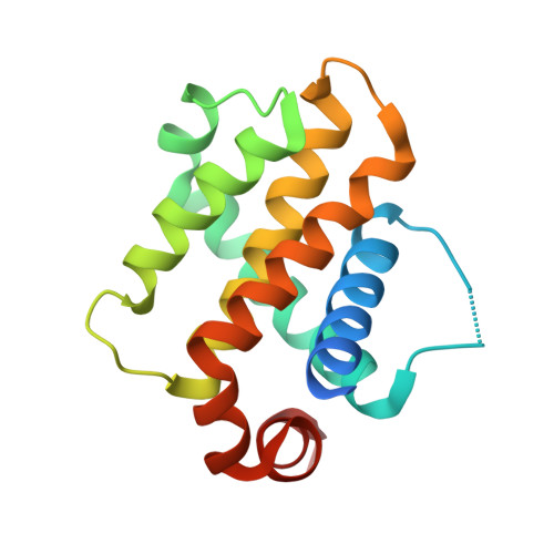
    BCL-2 family proteins are high-priority cancer targets whose structures provide essential blueprints for drug design. Whereas numerous structures of anti-apoptotic BCL-2 protein complexes with α-helical BH3 peptides have been reported, the corresponding panel of apo structures remains incomplete. Here, we report the crystal structure of apo BFL-1 at 1.69-Å resolution, revealing similarities and key differences among unliganded anti-apoptotic proteins. Unlike all other BCL-2 proteins, apo BFL-1 contains a surface-accessible cysteine within its BH3-binding groove, allowing for selective covalent targeting by a NOXA BH3-based stapled peptide inhibitor. The crystal structure of this complex demonstrated the sulfhydryl bond and fortuitous interactions between the acrylamide-bearing moiety and a newly formed hydrophobic cavity. Comparison of the apo and BH3-liganded structures further revealed an induced conformational change. The two BFL-1 structures expand our understanding of the surface landscapes available for therapeutic targeting so that the apoptotic blockades of BFL-1-dependent cancers can be overcome.


  • Organizational Affiliation

    Department of Pediatric Oncology, Dana-Farber Cancer Institute, Boston, MA 02215, USA; Linde Program in Cancer Chemical Biology, Dana-Farber Cancer Institute, Boston, MA 02215, USA.


Macromolecules
Find similar proteins by:  (by identity cutoff)  |  3D Structure
Entity ID: 1
MoleculeChains Sequence LengthOrganismDetailsImage
Bcl-2-related protein A1161Homo sapiensMutation(s): 2 
Gene Names: BCL2A1BCL2L5BFL1GRSHBPA1
UniProt & NIH Common Fund Data Resources
Find proteins for Q16548 (Homo sapiens)
Explore Q16548 
Go to UniProtKB:  Q16548
PHAROS:  Q16548
GTEx:  ENSG00000140379 
Entity Groups  
Sequence Clusters30% Identity50% Identity70% Identity90% Identity95% Identity100% Identity
UniProt GroupQ16548
Sequence Annotations
Expand
  • Reference Sequence
Small Molecules
Ligands 1 Unique
IDChains Name / Formula / InChI Key2D Diagram3D Interactions
CAD
Query on CAD

Download Ideal Coordinates CCD File 
B [auth A]CACODYLIC ACID
C2 H7 As O2
OGGXGZAMXPVRFZ-UHFFFAOYSA-N
Experimental Data & Validation

Experimental Data

  • Method: X-RAY DIFFRACTION
  • Resolution: 1.69 Å
  • R-Value Free: 0.233 
  • R-Value Work: 0.195 
  • R-Value Observed: 0.197 
  • Space Group: P 1 21 1
Unit Cell:
Length ( Å )Angle ( ˚ )
a = 39.51α = 90
b = 43.09β = 104.15
c = 43.44γ = 90
Software Package:
Software NamePurpose
PHENIXrefinement
xia2data scaling
PHASERphasing
PDB_EXTRACTdata extraction
XDSdata reduction

Structure Validation

View Full Validation Report


Entry History 

Deposition Data

Revision History  (Full details and data files)

  • Version 1.0: 2018-01-17
    Type: Initial release
  • Version 1.1: 2023-10-04
    Changes: Data collection, Database references, Refinement description