5XCO | pdb_00005xco

Crystal structure of human K-Ras G12D Mutant in complex with GDP and Cyclic Inhibitory Peptide


Experimental Data Snapshot

  • Method: X-RAY DIFFRACTION
  • Resolution: 1.25 Å
  • R-Value Free: 
    0.194 (Depositor), 0.199 (DCC) 
  • R-Value Work: 
    0.171 (Depositor), 0.179 (DCC) 
  • R-Value Observed: 
    0.172 (Depositor) 

Starting Model: experimental
View more details

wwPDB Validation 3D Report Full Report

Validation slider image for 5XCO

Ligand Structure Quality Assessment 


This is version 1.3 of the entry. See complete history

Literature

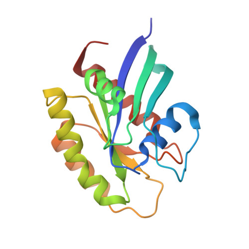
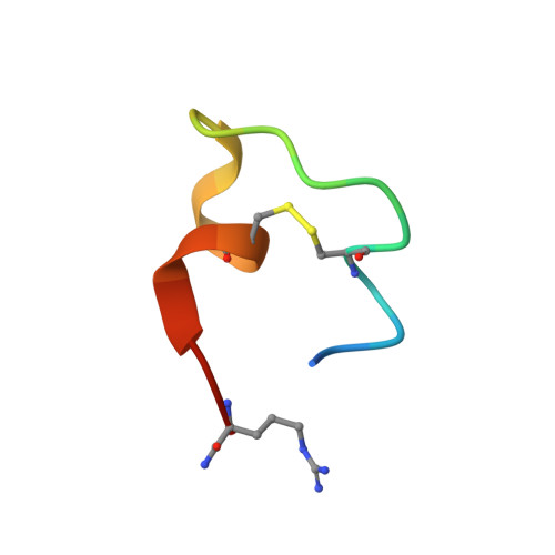
Crystal Structure of a Human K-Ras G12D Mutant in Complex with GDP and the Cyclic Inhibitory Peptide KRpep-2d

Sogabe, S.Kamada, Y.Miwa, M.Niida, A.Sameshima, T.Kamaura, M.Yonemori, K.Sasaki, S.Sakamoto, J.Sakamoto, K.

(2017) ACS Med Chem Lett 8: 732-736

  • DOI: https://doi.org/10.1021/acsmedchemlett.7b00128
  • Primary Citation Related Structures: 
    5XCO

  • PubMed Abstract: 

    The Ras proteins play roles in cell differentiation, proliferation, and survival. Aberrant signaling through Ras-mediated pathways in tumor cells occurs as a result of several types of mutational damage, which most frequently affects the amino acids G12, G13, and Q61. Recently, KRpep-2d was identified as a K-Ras(G12D) selective inhibitory peptide against the G12D mutant of K-Ras, which is a key member of the Ras protein family and an attractive cancer therapeutic target. In this study, the crystal structure of the human K-Ras(G12D) mutant was determined in complex with GDP and KRpep-2d at 1.25 Å resolution. This structure revealed that the peptide binds near Switch II and allosterically blocks protein-protein interactions with the guanine nucleotide exchange factor. This discovery of a unique binding pocket provides valuable information that will facilitate the design of direct Ras inhibitors.


  • Organizational Affiliation
    • Pharmaceutical Research Division, Takeda Pharmaceutical Company Limited, 26-1, Muraoka-Higashi 2-chome, Fujisawa, Kanagawa 251-8555, Japan.

Macromolecule Content 

  • Total Structure Weight: 22.53 kDa 
  • Atom Count: 1,780 
  • Modeled Residue Count: 191 
  • Deposited Residue Count: 192 
  • Unique protein chains: 2

Macromolecules

Find similar proteins by:|  3D Structure
Entity ID: 1
MoleculeChains  Sequence LengthOrganismDetailsImage
GTPase KRas171Homo sapiensMutation(s): 1 
Gene Names: KRASKRAS2RASK2
EC: 3.6.5.2
UniProt & NIH Common Fund Data Resources
Find proteins for P01116 (Homo sapiens)
Explore P01116 
Go to UniProtKB:  P01116
PHAROS:  P01116
GTEx:  ENSG00000133703 
Entity Groups
Sequence Clusters30% Identity50% Identity70% Identity90% Identity95% Identity100% Identity
UniProt GroupP01116
Sequence Annotations
Expand
Reference Sequence
Find similar proteins by:  Sequence   |   3D Structure  
Entity ID: 2
MoleculeChains  Sequence LengthOrganismDetailsImage
ACE-ARG-ARG-ARG-ARG-CYS-PRO-LEU-TYR-ILE-SER-TYR-ASP-PRO-VAL-CYS-ARG-ARG-ARG-ARG-NH221Escherichia phage T7Mutation(s): 0 

Small Molecules

Experimental Data & Validation

Experimental Data

  • Method: X-RAY DIFFRACTION
  • Resolution: 1.25 Å
  • R-Value Free:  0.194 (Depositor), 0.199 (DCC) 
  • R-Value Work:  0.171 (Depositor), 0.179 (DCC) 
  • R-Value Observed: 0.172 (Depositor) 
Space Group: P 31 2 1
Unit Cell:
Length ( Å )Angle ( ˚ )
a = 51.529α = 90
b = 51.529β = 90
c = 129.741γ = 120
Software Package:
Software NamePurpose
REFMACrefinement
HKL-2000data reduction
SCALEPACKdata scaling
PHASERphasing

Structure Validation

View Full Validation Report


Ligand Structure Quality Assessment 


Entry History 

Deposition Data

Revision History  (Full details and data files)

  • Version 1.0: 2017-05-24
    Type: Initial release
  • Version 1.1: 2017-08-09
    Changes: Database references
  • Version 1.2: 2023-11-22
    Changes: Data collection, Database references, Refinement description
  • Version 1.3: 2024-10-23
    Changes: Structure summary