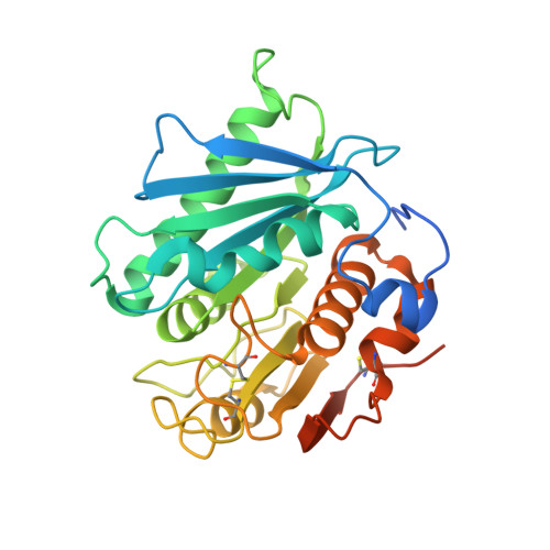
5XJH

Crystal structure of PETase from Ideonella sakaiensis


Experimental Data Snapshot

  • Method: X-RAY DIFFRACTION
  • Resolution: 1.54 Å
  • R-Value Free: 0.192 
  • R-Value Work: 0.160 
  • R-Value Observed: 0.161 

Starting Model: experimental
View more details

wwPDB Validation   3D Report Full Report


This is version 1.3 of the entry. See complete history


Literature

Structural insight into molecular mechanism of poly(ethylene terephthalate) degradation.

Joo, S.Cho, I.J.Seo, H.Son, H.F.Sagong, H.-Y.Shin, T.J.Choi, S.Y.Lee, S.Y.Kim, K.-J.

(2018) Nat Commun 9: 382-382

  • DOI: https://doi.org/10.1038/s41467-018-02881-1
  • Primary Citation of Related Structures:  
    5XJH, 5YNS

  • PubMed Abstract: 

    Plastics, including poly(ethylene terephthalate) (PET), possess many desirable characteristics and thus are widely used in daily life. However, non-biodegradability, once thought to be an advantage offered by plastics, is causing major environmental problem. Recently, a PET-degrading bacterium, Ideonella sakaiensis, was identified and suggested for possible use in degradation and/or recycling of PET. However, the molecular mechanism of PET degradation is not known. Here we report the crystal structure of I. sakaiensis PETase (IsPETase) at 1.5 Å resolution. IsPETase has a Ser-His-Asp catalytic triad at its active site and contains an optimal substrate binding site to accommodate four monohydroxyethyl terephthalate (MHET) moieties of PET. Based on structural and site-directed mutagenesis experiments, the detailed process of PET degradation into MHET, terephthalic acid, and ethylene glycol is suggested. Moreover, other PETase candidates potentially having high PET-degrading activities are suggested based on phylogenetic tree analysis of 69 PETase-like proteins.


  • Organizational Affiliation

    School of Life Sciences (KNU Creative BioResearch Group), KNU Institute for Microorganisms, Kyungpook National University, Daehak-ro 80, Buk-gu, Daegu, 41566, Republic of Korea.


Macromolecules
Find similar proteins by:  (by identity cutoff)  |  3D Structure
Entity ID: 1
MoleculeChains Sequence LengthOrganismDetailsImage
Poly(ethylene terephthalate) hydrolase300Piscinibacter sakaiensisMutation(s): 0 
Gene Names: ISF6_4831
EC: 3.1.1.101
UniProt
Find proteins for A0A0K8P6T7 (Piscinibacter sakaiensis)
Explore A0A0K8P6T7 
Go to UniProtKB:  A0A0K8P6T7
Entity Groups  
Sequence Clusters30% Identity50% Identity70% Identity90% Identity95% Identity100% Identity
UniProt GroupA0A0K8P6T7
Sequence Annotations
Expand
  • Reference Sequence
Experimental Data & Validation

Experimental Data

  • Method: X-RAY DIFFRACTION
  • Resolution: 1.54 Å
  • R-Value Free: 0.192 
  • R-Value Work: 0.160 
  • R-Value Observed: 0.161 
  • Space Group: P 21 21 21
Unit Cell:
Length ( Å )Angle ( ˚ )
a = 43.475α = 90
b = 50.395β = 90
c = 129.49γ = 90
Software Package:
Software NamePurpose
REFMACrefinement
HKL-2000data processing

Structure Validation

View Full Validation Report


Entry History 

Deposition Data

Revision History  (Full details and data files)

  • Version 1.0: 2018-02-14
    Type: Initial release
  • Version 1.1: 2020-09-16
    Changes: Structure summary
  • Version 1.2: 2023-11-22
    Changes: Data collection, Database references, Refinement description
  • Version 1.3: 2024-10-30
    Changes: Structure summary