Atomic structures of FUS LC domain segments reveal bases for reversible amyloid fibril formation.
Luo, F., Gui, X., Zhou, H., Gu, J., Li, Y., Liu, X., Zhao, M., Li, D., Li, X., Liu, C.(2018) Nat Struct Mol Biol 25: 341-346
- PubMed: 29610493 Search on PubMed
- DOI: https://doi.org/10.1038/s41594-018-0050-8
- Primary Citation Related Structures:
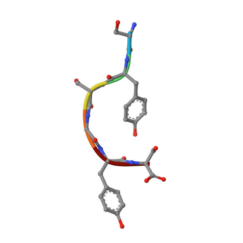
5XRR, 5XSG - PubMed Abstract:
Thermostable cross-β structures are characteristic of pathological amyloid fibrils, but these structures cannot explain the reversible nature of fibrils formed by RNA-binding proteins such as fused in sarcoma (FUS), involved in RNA granule assembly. Here, we find that two tandem (S/G)Y(S/G) motifs of the human FUS low-complexity domain (FUS LC) form reversible fibrils in a temperature- and phosphorylation-dependent manner. We named these motifs reversible amyloid cores, or RAC1 and RAC2, and determined their atomic structures in fibrillar forms, using microelectron and X-ray diffraction techniques. The RAC1 structure features an ordered-coil fibril spine rather than the extended β-strand typical of amyloids. Ser42, a phosphorylation site of FUS, is critical in the maintenance of the ordered-coil structure, which explains how phosphorylation controls fibril formation. The RAC2 structure shows a labile fibril spine with a wet interface. These structures illuminate the mechanism of reversible fibril formation and dynamic assembly of RNA granules.
- Interdisciplinary Research Center on Biology and Chemistry, Shanghai Institute of Organic Chemistry, Chinese Academy of Sciences, Shanghai, China.
Organizational Affiliation: