5YGX | pdb_00005ygx

Structure of BACE1 in complex with N-(3-((4R,5R,6S)-2-amino-6-(1,1-difluoroethyl)-5-fluoro-4-methyl-5,6-dihydro-4H-1,3-oxazin-4-yl)-4-fluorophenyl)-5-(fluoromethoxy)pyrazine-2-carboxamide


Experimental Data Snapshot

  • Method: X-RAY DIFFRACTION
  • Resolution: 2.20 Å
  • R-Value Free: 
    0.207 (Depositor), 0.211 (DCC) 
  • R-Value Work: 
    0.178 (Depositor), 0.184 (DCC) 
  • R-Value Observed: 
    0.180 (Depositor) 

Starting Model: experimental
View more details

wwPDB Validation 3D Report Full Report

Validation slider image for 5YGX

Ligand Structure Quality Assessment 


This is version 1.2 of the entry. See complete history

Literature

Discovery of Potent and Centrally Active 6-Substituted 5-Fluoro-1,3-dihydro-oxazine beta-Secretase (BACE1) Inhibitors via Active Conformation Stabilization

Nakahara, K.Fuchino, K.Komano, K.Asada, N.Tadano, G.Hasegawa, T.Yamamoto, T.Sako, Y.Ogawa, M.Unemura, C.Hosono, M.Ito, H.Sakaguchi, G.Ando, S.Ohnishi, S.Kido, Y.Fukushima, T.Dhuyvetter, D.Borghys, H.Gijsen, H.J.M.Yamano, Y.Iso, Y.Kusakabe, K.I.

(2018) J Med Chem 61: 5525-5546

  • DOI: https://doi.org/10.1021/acs.jmedchem.8b00011
  • Primary Citation Related Structures: 
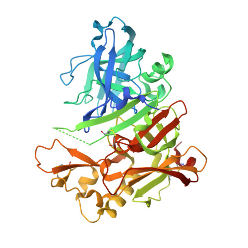
    5YGX

  • PubMed Abstract: 

    β-Secretase (BACE1) has an essential role in the production of amyloid β peptides that accumulate in patients with Alzheimer's disease (AD). Thus, inhibition of BACE1 is considered to be a disease-modifying approach for the treatment of AD. Our hit-to-lead efforts led to a cellular potent 1,3-dihydro-oxazine 6, which however inhibited hERG and showed high P-gp efflux. The close analogue of 5-fluoro-oxazine 8 reduced P-gp efflux; further introduction of electron withdrawing groups at the 6-position improved potency and also mitigated P-gp efflux and hERG inhibition. Changing to a pyrazine followed by optimization of substituents on both the oxazine and the pyrazine culminated in 24 with robust Aβ reduction in vivo at low doses as well as reduced CYP2D6 inhibition. On the basis of the X-ray analysis and the QM calculation of given dihydro-oxazines, we reasoned that the substituents at the 6-position as well as the 5-fluorine on the oxazine would stabilize a bioactive conformation to increase potency.

Macromolecule Content 

  • Total Structure Weight: 47.64 kDa 
  • Atom Count: 3,292 
  • Modeled Residue Count: 381 
  • Deposited Residue Count: 416 
  • Unique protein chains: 1

Macromolecules

Find similar proteins by:|  3D Structure
Entity ID: 1
MoleculeChains  Sequence LengthOrganismDetailsImage
Beta-secretase 1416Homo sapiensMutation(s): 0 
Gene Names: BACE1BACEKIAA1149
EC: 3.4.23.46
UniProt & NIH Common Fund Data Resources
Find proteins for P56817 (Homo sapiens)
Explore P56817 
Go to UniProtKB:  P56817
PHAROS:  P56817
GTEx:  ENSG00000186318 
Entity Groups
Sequence Clusters30% Identity50% Identity70% Identity90% Identity95% Identity100% Identity
UniProt GroupP56817
Sequence Annotations
Expand
Reference Sequence

Small Molecules

Ligands 4 Unique
IDChains Name / Formula / InChI Key2D Diagram3D Interactions
0B5
(Subject of Investigation/LOI)

Query on 0B5


Download:Ideal Coordinates CCD File
J [auth A]~{N}-[3-[(4~{R},5~{R},6~{S})-2-azanyl-6-[1,1-bis(fluoranyl)ethyl]-5-fluoranyl-4-methyl-5,6-dihydro-1,3-oxazin-4-yl]-4-fluoranyl-phenyl]-5-(fluoranylmethoxy)pyrazine-2-carboxamide
C19 H18 F5 N5 O3
DKIWJWSKVCKJPV-RLFYNMQTSA-N
IOD

Query on IOD


Download:Ideal Coordinates CCD File
B [auth A],
C [auth A]
IODIDE ION
I
XMBWDFGMSWQBCA-UHFFFAOYSA-M
GOL

Query on GOL


Download:Ideal Coordinates CCD File
D [auth A],
E [auth A],
F [auth A],
G [auth A],
H [auth A]
GLYCEROL
C3 H8 O3
PEDCQBHIVMGVHV-UHFFFAOYSA-N
DMS

Query on DMS


Download:Ideal Coordinates CCD File
I [auth A]DIMETHYL SULFOXIDE
C2 H6 O S
IAZDPXIOMUYVGZ-UHFFFAOYSA-N
Binding Affinity Annotations 
IDSourceBinding Affinity
0B5 BindingDB:  5YGX IC50: min: 1.9, max: 8.7 (nM) from 2 assay(s)

Experimental Data & Validation

Experimental Data

  • Method: X-RAY DIFFRACTION
  • Resolution: 2.20 Å
  • R-Value Free:  0.207 (Depositor), 0.211 (DCC) 
  • R-Value Work:  0.178 (Depositor), 0.184 (DCC) 
  • R-Value Observed: 0.180 (Depositor) 
Space Group: P 61 2 2
Unit Cell:
Length ( Å )Angle ( ˚ )
a = 101.55α = 90
b = 101.55β = 90
c = 170.811γ = 120
Software Package:
Software NamePurpose
MOSFLMdata reduction
SCALAdata scaling
MOLREPphasing
REFMACrefinement
PDB_EXTRACTdata extraction

Structure Validation

View Full Validation Report


Ligand Structure Quality Assessment 


Entry History 

Revision History  (Full details and data files)

  • Version 1.0: 2018-08-08
    Type: Initial release
  • Version 1.1: 2023-11-22
    Changes: Data collection, Database references, Refinement description
  • Version 1.2: 2024-11-20
    Changes: Structure summary