5ZIA | pdb_00005zia

Crystal structure of human anti-tau antibody CBTAU-24.1 in complex with its phosphorylated tau peptide

  • Classification: IMMUNE SYSTEM
  • Organism(s): Homo sapiens
  • Expression System: Homo sapiens
  • Mutation(s): No 

  • Deposited: 2018-03-14 Released: 2018-10-31 
  • Deposition Author(s): Zhang, H., Wilson, I.A.
  • Funding Organization(s): National Institutes of Health/National Institute of Arthritis and Musculoskeletal and Skin Diseases (NIH/NIAMS)

Experimental Data Snapshot

  • Method: X-RAY DIFFRACTION
  • Resolution: 2.60 Å
  • R-Value Free: 
    0.249 (Depositor), 0.250 (DCC) 
  • R-Value Work: 
    0.200 (Depositor), 0.203 (DCC) 
  • R-Value Observed: 
    0.202 (Depositor) 

Starting Model: experimental
View more details

wwPDB Validation 3D Report Full Report

Validation slider image for 5ZIA

This is version 1.5 of the entry. See complete history

Literature

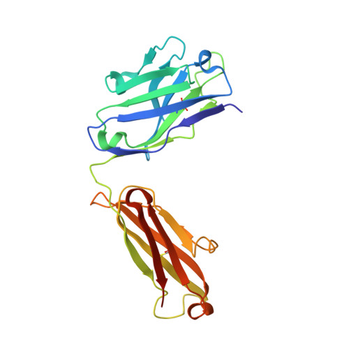
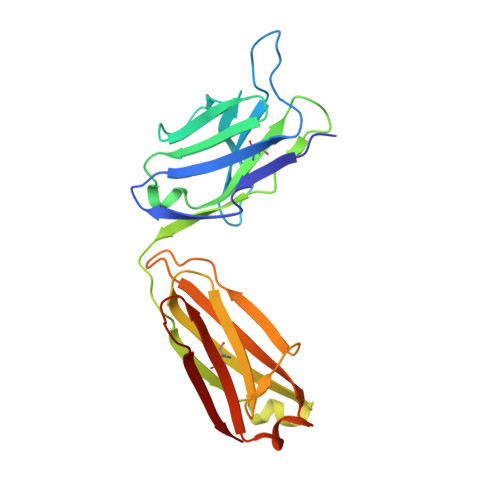
Structural Basis for Recognition of a Unique Epitope by a Human Anti-tau Antibody.

Zhang, H.Zhu, X.Pascual, G.Wadia, J.S.Keogh, E.Hoozemans, J.J.Siregar, B.Inganas, H.Stoop, E.J.M.Goudsmit, J.Apetri, A.Koudstaal, W.Wilson, I.A.

(2018) Structure 26: 1626-1634.e4

  • DOI: https://doi.org/10.1016/j.str.2018.08.012
  • Primary Citation Related Structures: 
    5ZIA

  • PubMed Abstract: 

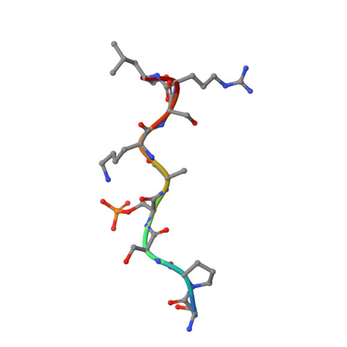
    Aggregation of the hyperphosphorylated protein tau into neurofibrillary tangles and neuropil threads is a hallmark of Alzheimer disease (AD). Identification and characterization of the epitopes recognized by anti-tau antibodies might shed light on the molecular mechanisms of AD pathogenesis. Here we report on the biochemical and structural characterization of a tau-specific monoclonal antibody CBTAU-24.1, which was isolated from the human memory B cell repertoire. Immunohistochemical staining with CBTAU-24.1 specifically detects pathological tau structures in AD brain samples. The crystal structure of CBTAU-24.1 Fab with a phosphorylated tau peptide revealed recognition of a unique epitope (Ser235-Leu243) in the tau proline-rich domain. Interestingly, the antibody can bind tau regardless of phosphorylation state of its epitope region and also recognizes both monomeric and paired helical filament tau irrespective of phosphorylation status. This human anti-tau antibody and its unique epitope may aid in development of diagnostics and/or therapeutic AD strategies.


  • Organizational Affiliation
    • Department of Integrative Structural and Computational Biology, The Scripps Research Institute, La Jolla, CA 92037, USA; Beijing Synchrotron Radiation Facility, Institute of High Energy Physics, Chinese Academy of Sciences, Beijing 100049, People's Republic of China.

Macromolecule Content 

  • Total Structure Weight: 297.45 kDa 
  • Atom Count: 20,828 
  • Modeled Residue Count: 2,675 
  • Deposited Residue Count: 2,730 
  • Unique protein chains: 3

Macromolecules

Find similar proteins by:|  3D Structure
Entity ID: 1
MoleculeChains  Sequence LengthOrganismDetailsImage
Heavy chain of CBTAU-24.1 antibody226Homo sapiensMutation(s): 0 
Find similar proteins by:|  3D Structure
Entity ID: 2
MoleculeChains  Sequence LengthOrganismDetailsImage
Light chain (kappa) of CBTAU-24.1 antibody220Homo sapiensMutation(s): 0 
Find similar proteins by:  Sequence   |   3D Structure  
Entity ID: 3
MoleculeChains  Sequence LengthOrganismDetailsImage
phosphorylated tau peptide9Homo sapiensMutation(s): 0 
UniProt & NIH Common Fund Data Resources
Find proteins for P10636 (Homo sapiens)
Explore P10636 
Go to UniProtKB:  P10636
PHAROS:  P10636
GTEx:  ENSG00000186868 
Entity Groups
UniProt GroupP10636
Sequence Annotations
Expand
Reference Sequence

Small Molecules

Modified Residues  1 Unique
IDChains TypeFormula2D DiagramParent
SEP
Query on SEP
C [auth R]
F [auth C]
I [auth F]
L [auth J]
O [auth N]
C [auth R],
F [auth C],
I [auth F],
L [auth J],
O [auth N],
R [auth Q]
L-PEPTIDE LINKINGC3 H8 N O6 PSER

Experimental Data & Validation

Experimental Data

  • Method: X-RAY DIFFRACTION
  • Resolution: 2.60 Å
  • R-Value Free:  0.249 (Depositor), 0.250 (DCC) 
  • R-Value Work:  0.200 (Depositor), 0.203 (DCC) 
  • R-Value Observed: 0.202 (Depositor) 
Space Group: P 1
Unit Cell:
Length ( Å )Angle ( ˚ )
a = 86.353α = 62.16
b = 107.811β = 71.02
c = 107.931γ = 90.77
Software Package:
Software NamePurpose
PHENIXrefinement
HKL-2000data reduction
HKL-2000data scaling
PHASERphasing

Structure Validation

View Full Validation Report


Entry History 

& Funding Information

Deposition Data


Funding OrganizationLocationGrant Number
National Institutes of Health/National Institute of Arthritis and Musculoskeletal and Skin Diseases (NIH/NIAMS)United StatesP41GM103393

Revision History  (Full details and data files)

  • Version 1.0: 2018-10-31
    Type: Initial release
  • Version 1.1: 2018-12-26
    Changes: Data collection, Database references
  • Version 1.2: 2019-12-25
    Changes: Derived calculations
  • Version 1.3: 2022-03-23
    Changes: Author supporting evidence, Database references
  • Version 1.4: 2023-11-22
    Changes: Data collection, Refinement description
  • Version 1.5: 2024-11-20
    Changes: Structure summary