5ZMD | pdb_00005zmd

Crystal structure of FTO in complex with m6dA modified ssDNA


Experimental Data Snapshot

  • Method: X-RAY DIFFRACTION
  • Resolution: 3.30 Å
  • R-Value Free: 
    0.292 (Depositor), 0.295 (DCC) 
  • R-Value Work: 
    0.271 (Depositor), 0.275 (DCC) 
  • R-Value Observed: 
    0.272 (Depositor) 

Starting Model: experimental
View more details

wwPDB Validation 3D Report Full Report

Validation slider image for 5ZMD

This is version 1.2 of the entry. See complete history

Literature

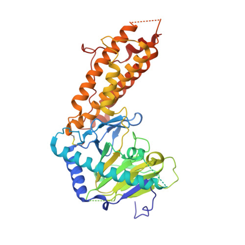
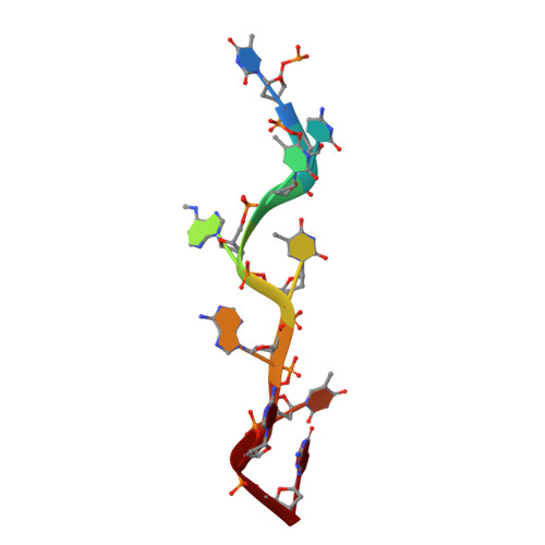
Structural insights into FTO's catalytic mechanism for the demethylation of multiple RNA substrates.

Zhang, X.Wei, L.H.Wang, Y.Xiao, Y.Liu, J.Zhang, W.Yan, N.Amu, G.Tang, X.Zhang, L.Jia, G.

(2019) Proc Natl Acad Sci U S A 116: 2919-2924

  • DOI: https://doi.org/10.1073/pnas.1820574116
  • Primary Citation Related Structures: 
    5ZMD

  • PubMed Abstract: 

    FTO demethylates internal N 6 -methyladenosine (m 6 A) and N 6 ,2'- O -dimethyladenosine (m 6 A m ; at the cap +1 position) in mRNA, m 6 A and m 6 A m in snRNA, and N 1 -methyladenosine (m 1 A) in tRNA in vivo, and in vitro evidence supports that it can also demethylate N 6 -methyldeoxyadenosine (6mA), 3-methylthymine (3mT), and 3-methyluracil (m 3 U). However, it remains unclear how FTO variously recognizes and catalyzes these diverse substrates. Here we demonstrate-in vitro and in vivo-that FTO has extensive demethylation enzymatic activity on both internal m 6 A and cap m 6 A m Considering that 6mA, m 6 A, and m 6 A m all share the same nucleobase, we present a crystal structure of human FTO bound to 6mA-modified ssDNA, revealing the molecular basis of the catalytic demethylation of FTO toward multiple RNA substrates. We discovered that ( i ) N 6 -methyladenine is the most favorable nucleobase substrate of FTO, ( ii ) FTO displays the same demethylation activity toward internal m 6 A and m 6 A m in the same RNA sequence, suggesting that the substrate specificity of FTO primarily results from the interaction of residues in the catalytic pocket with the nucleobase (rather than the ribose ring), and ( iii ) the sequence and the tertiary structure of RNA can affect the catalytic activity of FTO. Our findings provide a structural basis for understanding the catalytic mechanism through which FTO demethylates its multiple substrates and pave the way forward for the structure-guided design of selective chemicals for functional studies and potential therapeutic applications.


  • Organizational Affiliation
    • Synthetic and Functional Biomolecules Center, Beijing National Laboratory for Molecular Sciences, Key Laboratory of Bioorganic Chemistry and Molecular Engineering of Ministry of Education, College of Chemistry and Molecular Engineering, Peking University, Beijing 100871, China.

Macromolecule Content 

  • Total Structure Weight: 224.94 kDa 
  • Atom Count: 13,828 
  • Modeled Residue Count: 1,631 
  • Deposited Residue Count: 1,888 
  • Unique protein chains: 1
  • Unique nucleic acid chains: 1

Macromolecules


Find similar proteins by:|  3D Structure
Entity ID: 1
MoleculeChains  Sequence LengthOrganismDetailsImage
Alpha-ketoglutarate-dependent dioxygenase FTO
A, C, E, G
463Homo sapiensMutation(s): 2 
Gene Names: FTOKIAA1752
EC: 1.14.11 (PDB Primary Data), 1.14.11.53 (UniProt)
UniProt & NIH Common Fund Data Resources
Find proteins for Q9C0B1 (Homo sapiens)
Explore Q9C0B1 
Go to UniProtKB:  Q9C0B1
PHAROS:  Q9C0B1
GTEx:  ENSG00000140718 
Entity Groups
Sequence Clusters30% Identity50% Identity70% Identity90% Identity95% Identity100% Identity
UniProt GroupQ9C0B1
Sequence Annotations
Expand
Reference Sequence
Find similar nucleic acids by:  Sequence
Entity ID: 2
MoleculeChains LengthOrganismImage
DNA (5'-D(P*TP*CP*TP*(6MA)P*TP*AP*TP*CP*G)-3')
B, D, F, H
9DNA launch vector pDE-GFP2
Sequence Annotations
Expand
Reference Sequence

Small Molecules

Ligands 2 Unique
IDChains Name / Formula / InChI Key2D Diagram3D Interactions
OGA

Query on OGA


Download:Ideal Coordinates CCD File
J [auth A],
L [auth C],
N [auth E],
P [auth G]
N-OXALYLGLYCINE
C4 H5 N O5
BIMZLRFONYSTPT-UHFFFAOYSA-N
MN

Query on MN


Download:Ideal Coordinates CCD File
I [auth A],
K [auth C],
M [auth E],
O [auth G]
MANGANESE (II) ION
Mn
WAEMQWOKJMHJLA-UHFFFAOYSA-N
Binding Affinity Annotations 
IDSourceBinding Affinity
OGA BindingDB:  5ZMD IC50: min: 3.65e+4, max: 4.40e+4 (nM) from 2 assay(s)

Experimental Data & Validation

Experimental Data

  • Method: X-RAY DIFFRACTION
  • Resolution: 3.30 Å
  • R-Value Free:  0.292 (Depositor), 0.295 (DCC) 
  • R-Value Work:  0.271 (Depositor), 0.275 (DCC) 
  • R-Value Observed: 0.272 (Depositor) 
Space Group: I 21 21 21
Unit Cell:
Length ( Å )Angle ( ˚ )
a = 122.736α = 90
b = 160.033β = 90
c = 276.756γ = 90
Software Package:
Software NamePurpose
PHENIXrefinement
HKL-3000data reduction
HKL-3000data scaling
PHASERphasing

Structure Validation

View Full Validation Report


Entry History 

& Funding Information

Deposition Data


Funding OrganizationLocationGrant Number
National Natural Science Foundation of ChinaChina21432002
National Natural Science Foundation of ChinaChina21722802

Revision History  (Full details and data files)

  • Version 1.0: 2019-04-10
    Type: Initial release
  • Version 1.1: 2023-11-22
    Changes: Data collection, Database references, Derived calculations, Refinement description
  • Version 1.2: 2024-10-30
    Changes: Structure summary