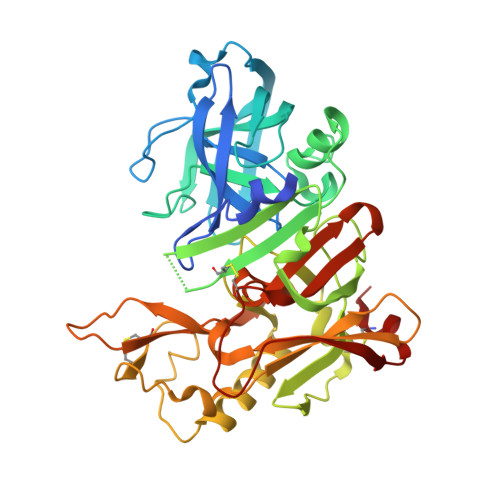
5QD2

Crystal structure of BACE complex with BMC017


Experimental Data Snapshot

  • Method: X-RAY DIFFRACTION
  • Resolution: 2.50 Å
  • R-Value Free: 0.205 
  • R-Value Work: 0.157 
  • R-Value Observed: 0.160 

wwPDB Validation   3D Report Full Report


Ligand Structure Quality Assessment 


This is version 1.2 of the entry. See complete history


Literature

D3R grand challenge 4: blind prediction of protein-ligand poses, affinity rankings, and relative binding free energies.

Parks, C.D.Gaieb, Z.Chiu, M.Yang, H.Shao, C.Walters, W.P.Jansen, J.M.McGaughey, G.Lewis, R.A.Bembenek, S.D.Ameriks, M.K.Mirzadegan, T.Burley, S.K.Amaro, R.E.Gilson, M.K.

(2020) J Comput Aided Mol Des 34: 99-119

  • DOI: https://doi.org/10.1007/s10822-020-00289-y
  • Primary Citation of Related Structures:  
    5QCO, 5QCP, 5QCQ, 5QCR, 5QCS, 5QCT, 5QCU, 5QCV, 5QCW, 5QCX, 5QCY, 5QCZ, 5QD0, 5QD1, 5QD2, 5QD3, 5QD4, 5QD5, 5QD6, 5QD7, 5QD8, 5QD9, 5QDA, 5QDB, 5QDC, 5QDD

  • PubMed Abstract: 

    The Drug Design Data Resource (D3R) aims to identify best practice methods for computer aided drug design through blinded ligand pose prediction and affinity challenges. Herein, we report on the results of Grand Challenge 4 (GC4). GC4 focused on proteins beta secretase 1 and Cathepsin S, and was run in an analogous manner to prior challenges. In Stage 1, participant ability to predict the pose and affinity of BACE1 ligands were assessed. Following the completion of Stage 1, all BACE1 co-crystal structures were released, and Stage 2 tested affinity rankings with co-crystal structures. We provide an analysis of the results and discuss insights into determined best practice methods.


  • Organizational Affiliation

    Drug Design Data Resource, University of California, San Diego, La Jolla, CA, 92093, USA.


Macromolecules
Find similar proteins by:  (by identity cutoff)  |  3D Structure
Entity ID: 1
MoleculeChains Sequence LengthOrganismDetailsImage
Beta-secretase 1
A, B, C
402Homo sapiensMutation(s): 0 
Gene Names: BACE1BACEKIAA1149
EC: 3.4.23.46
UniProt & NIH Common Fund Data Resources
Find proteins for P56817 (Homo sapiens)
Explore P56817 
Go to UniProtKB:  P56817
PHAROS:  P56817
GTEx:  ENSG00000186318 
Entity Groups  
Sequence Clusters30% Identity50% Identity70% Identity90% Identity95% Identity100% Identity
UniProt GroupP56817
Sequence Annotations
Expand
  • Reference Sequence
Small Molecules
Ligands 1 Unique
IDChains Name / Formula / InChI Key2D Diagram3D Interactions
E6J
Query on E6J

Download Ideal Coordinates CCD File 
D [auth A],
E [auth B],
F [auth C]
(4S)-4-[(1R)-1-hydroxy-2-({[3-(propan-2-yl)phenyl]methyl}amino)ethyl]-19-(methoxymethyl)-11,16-dioxa-3-azatricyclo[15.3.1.1~6,10~]docosa-1(21),6(22),7,9,17,19-hexaen-2-one
C33 H42 N2 O5
NGMBHSPURAEOBG-AJQTZOPKSA-N
Binding Affinity Annotations 
IDSourceBinding Affinity
E6J BindingDB:  5QD2 IC50: 72 (nM) from 1 assay(s)
Experimental Data & Validation

Experimental Data

  • Method: X-RAY DIFFRACTION
  • Resolution: 2.50 Å
  • R-Value Free: 0.205 
  • R-Value Work: 0.157 
  • R-Value Observed: 0.160 
  • Space Group: P 1 21 1
Unit Cell:
Length ( Å )Angle ( ˚ )
a = 82.077α = 90
b = 103.09β = 104.02
c = 100.342γ = 90
Software Package:
Software NamePurpose
PHENIXrefinement
CNXrefinement
CNXphasing
SCALEPACKdata scaling
HKL-2000date reduction
PDB_EXTRACTdata extraction

Structure Validation

View Full Validation Report


Ligand Structure Quality Assessment 


Entry History 

Deposition Data

Revision History  (Full details and data files)

  • Version 1.0: 2020-06-03
    Type: Initial release
  • Version 1.1: 2020-12-30
    Changes: Structure summary
  • Version 1.2: 2021-02-10
    Changes: Database references