5YKE

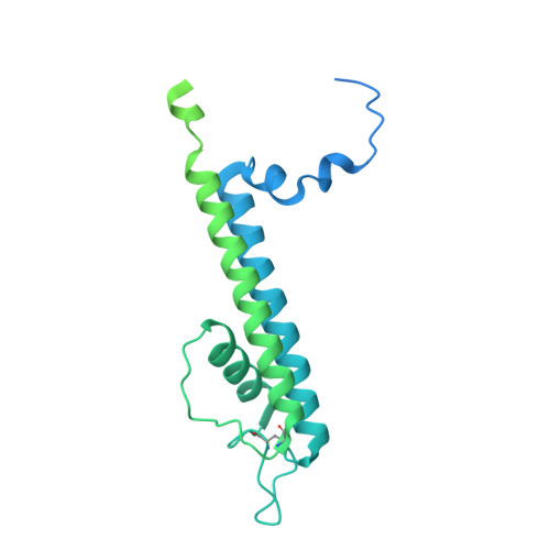
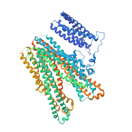
Structure of pancreatic ATP-sensitive potassium channel bound with glibenclamide and ATPgammaS (focused refinement on TM at 4.11A)


Experimental Data Snapshot

  • Method: ELECTRON MICROSCOPY
  • Resolution: 4.11 Å
  • Aggregation State: PARTICLE 
  • Reconstruction Method: SINGLE PARTICLE 

wwPDB Validation   3D Report Full Report


This is version 1.2 of the entry. See complete history


Literature

Ligand binding and conformational changes of SUR1 subunit in pancreatic ATP-sensitive potassium channels.

Wu, J.X.Ding, D.Wang, M.Kang, Y.Zeng, X.Chen, L.

(2018) Protein Cell 9: 553-567

  • DOI: https://doi.org/10.1007/s13238-018-0530-y
  • Primary Citation of Related Structures:  
    5YKE, 5YKF, 5YKG, 5YW7, 5YW8, 5YW9, 5YWA, 5YWB, 5YWC, 5YWD

  • PubMed Abstract: 

    ATP-sensitive potassium channels (K ATP ) are energy sensors on the plasma membrane. By sensing the intracellular ADP/ATP ratio of β-cells, pancreatic K ATP channels control insulin release and regulate metabolism at the whole body level. They are implicated in many metabolic disorders and diseases and are therefore important drug targets. Here, we present three structures of pancreatic K ATP channels solved by cryo-electron microscopy (cryo-EM), at resolutions ranging from 4.1 to 4.5 Å. These structures depict the binding site of the antidiabetic drug glibenclamide, indicate how Kir6.2 (inward-rectifying potassium channel 6.2) N-terminus participates in the coupling between the peripheral SUR1 (sulfonylurea receptor 1) subunit and the central Kir6.2 channel, reveal the binding mode of activating nucleotides, and suggest the mechanism of how Mg-ADP binding on nucleotide binding domains (NBDs) drives a conformational change of the SUR1 subunit.


  • Organizational Affiliation

    State Key Laboratory of Membrane Biology, Institute of Molecular Medicine, Peking University, Beijing Key Laboratory of Cardiometabolic Molecular Medicine, Beijing, 100871, China.


Macromolecules
Find similar proteins by:  (by identity cutoff)  |  3D Structure
Entity ID: 1
MoleculeChains Sequence LengthOrganismDetailsImage
ATP-sensitive inward rectifier potassium channel 11
A, C, E, G
390Mus musculusMutation(s): 0 
Gene Names: Kcnj11
Membrane Entity: Yes 
UniProt & NIH Common Fund Data Resources
Find proteins for Q61743 (Mus musculus)
Explore Q61743 
Go to UniProtKB:  Q61743
IMPC:  MGI:107501
Entity Groups  
Sequence Clusters30% Identity50% Identity70% Identity90% Identity95% Identity100% Identity
UniProt GroupQ61743
Sequence Annotations
Expand
  • Reference Sequence
Find similar proteins by:  (by identity cutoff)  |  3D Structure
Entity ID: 2
MoleculeChains Sequence LengthOrganismDetailsImage
ATP-binding cassette sub-family C member 8 isoform X2
B, D, F, H
1,582Mesocricetus auratusMutation(s): 0 
Gene Names: Abcc8
Membrane Entity: Yes 
UniProt
Find proteins for A0A1U7R319 (Mesocricetus auratus)
Explore A0A1U7R319 
Go to UniProtKB:  A0A1U7R319
Entity Groups  
Sequence Clusters30% Identity50% Identity70% Identity90% Identity95% Identity100% Identity
UniProt GroupA0A1U7R319
Sequence Annotations
Expand
  • Reference Sequence
Small Molecules
Binding Affinity Annotations 
IDSourceBinding Affinity
GBM BindingDB:  5YKE Ki: 130 (nM) from 1 assay(s)
IC50: min: 4.3, max: 220 (nM) from 2 assay(s)
Experimental Data & Validation

Experimental Data

  • Method: ELECTRON MICROSCOPY
  • Resolution: 4.11 Å
  • Aggregation State: PARTICLE 
  • Reconstruction Method: SINGLE PARTICLE 
EM Software:
TaskSoftware PackageVersion
RECONSTRUCTIONRELION2.0

Structure Validation

View Full Validation Report


Entry History & Funding Information

Deposition Data


Funding OrganizationLocationGrant Number
Ministry of Science and Technology of ChinaChina2016YFA0502004
National Natural Science Foundation of ChinaChina31622021
National Natural Science Foundation of ChinaChina31521062
China Postdoctoral Science FoundationChina2016M600856
China Postdoctoral Science FoundationChina2017T100014

Revision History  (Full details and data files)

  • Version 1.0: 2018-04-18
    Type: Initial release
  • Version 1.1: 2018-06-13
    Changes: Data collection, Database references
  • Version 1.2: 2024-11-20
    Changes: Data collection, Database references, Structure summary