6A6A

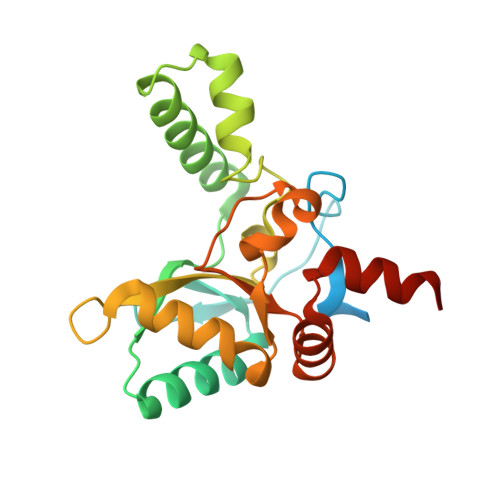
VanYB in complex with D-Alanine


Experimental Data Snapshot

  • Method: X-RAY DIFFRACTION
  • Resolution: 2.26 Å
  • R-Value Free: 0.276 
  • R-Value Work: 0.202 
  • R-Value Observed: 0.205 

Starting Model: experimental
View more details

wwPDB Validation   3D Report Full Report


Ligand Structure Quality Assessment 


This is version 1.1 of the entry. See complete history


Literature

Structural basis for the substrate recognition of peptidoglycan pentapeptides by Enterococcus faecalis VanYB.

Kim, H.S.Hahn, H.Kim, J.Jang, D.M.Lee, J.Y.Back, J.M.Im, H.N.Kim, H.Han, B.W.Suh, S.W.

(2018) Int J Biol Macromol 119: 335-344

  • DOI: https://doi.org/10.1016/j.ijbiomac.2018.07.081
  • Primary Citation of Related Structures:  
    5ZHF, 5ZHW, 6A6A

  • PubMed Abstract: 

    Vancomycin resistance in Enterococci and its transfer to methicillin-resistant Staphylococcus aureus are challenging problems in health care institutions worldwide. High-level vancomycin resistance is conferred by acquiring either transposable elements of the VanA or VanB type. Enterococcus faecalis VanY B in the VanB-type operon is a d,d-carboxypeptidase that recognizes the peptidyl-d-Ala 4 -d-Ala 5 extremity of peptidoglycan and hydrolyses the terminal d-Ala on the extracellular side of the cell wall, thereby increasing the level of glycopeptide antibiotics resistance. However, at the molecular level, it remains unclear how VanY B manipulates peptidoglycan peptides for vancomycin resistance. In this study, we have determined the crystal structures of E. faecalis VanY B in the d-Ala-d-Ala-bound, d-Ala-bound, and -unbound states. The interactions between VanY B and d-Ala-d-Ala observed in the crystal provide the molecular basis for the recognition of peptidoglycan substrates by VanY B . Moreover, comparisons with the related VanX and VanXY enzymes reveal distinct structural features of E. faecalis VanY B around the active-site cleft, thus shedding light on its unique substrate specificity. Our results could serve as the foundation for unravelling the molecular mechanism of vancomycin resistance and for developing novel antibiotics against the vancomycin-resistant Enterococcus species.


  • Organizational Affiliation

    Research Institute of Pharmaceutical Sciences, College of Pharmacy, Seoul National University, Seoul, Republic of Korea; Therapeutic Target Discovery Branch, Division of Precision Medicine, National Cancer Center, Goyang, Gyeonggi, Republic of Korea; Department of Chemistry, College of Natural Sciences, Seoul National University, Seoul, Republic of Korea. Electronic address: hskim@ncc.re.kr.


Macromolecules
Find similar proteins by:  (by identity cutoff)  |  3D Structure
Entity ID: 1
MoleculeChains Sequence LengthOrganismDetailsImage
D-alanyl-D-alanine carboxypeptidase
A, B
225Enterococcus faecalis V583Mutation(s): 0 
Gene Names: vanYBEF_2297
EC: 3.4.16.4 (PDB Primary Data), 3.4.17 (UniProt)
UniProt
Find proteins for Q47746 (Enterococcus faecalis (strain ATCC 700802 / V583))
Explore Q47746 
Go to UniProtKB:  Q47746
Entity Groups  
Sequence Clusters30% Identity50% Identity70% Identity90% Identity95% Identity100% Identity
UniProt GroupQ47746
Sequence Annotations
Expand
  • Reference Sequence
Small Molecules
Ligands 5 Unique
IDChains Name / Formula / InChI Key2D Diagram3D Interactions
PGE
Query on PGE

Download Ideal Coordinates CCD File 
H [auth A],
I [auth A],
O [auth B],
P [auth B]
TRIETHYLENE GLYCOL
C6 H14 O4
ZIBGPFATKBEMQZ-UHFFFAOYSA-N
GOL
Query on GOL

Download Ideal Coordinates CCD File 
D [auth A],
E [auth A],
K [auth B],
L [auth B]
GLYCEROL
C3 H8 O3
PEDCQBHIVMGVHV-UHFFFAOYSA-N
DAL (Subject of Investigation/LOI)
Query on DAL

Download Ideal Coordinates CCD File 
C [auth A],
J [auth B]
D-ALANINE
C3 H7 N O2
QNAYBMKLOCPYGJ-UWTATZPHSA-N
ZN
Query on ZN

Download Ideal Coordinates CCD File 
G [auth A],
N [auth B]
ZINC ION
Zn
PTFCDOFLOPIGGS-UHFFFAOYSA-N
ACT
Query on ACT

Download Ideal Coordinates CCD File 
F [auth A],
M [auth B]
ACETATE ION
C2 H3 O2
QTBSBXVTEAMEQO-UHFFFAOYSA-M
Experimental Data & Validation

Experimental Data

  • Method: X-RAY DIFFRACTION
  • Resolution: 2.26 Å
  • R-Value Free: 0.276 
  • R-Value Work: 0.202 
  • R-Value Observed: 0.205 
  • Space Group: C 1 2 1
Unit Cell:
Length ( Å )Angle ( ˚ )
a = 85.368α = 90
b = 40.83β = 109.08
c = 114.672γ = 90
Software Package:
Software NamePurpose
REFMACrefinement
HKL-2000data reduction
HKL-2000data scaling
MOLREPphasing

Structure Validation

View Full Validation Report


Ligand Structure Quality Assessment 


Entry History & Funding Information

Deposition Data


Funding OrganizationLocationGrant Number
Ministry of Science, ICT and Future PlanningKorea, Republic Of2013R1A2A1A05067303
Ministry of Science, ICT and Future PlanningKorea, Republic OfNRF-2011-0030001
Ministry of Science, ICT and Future PlanningKorea, Republic OfNRF-2013M3A6A4043695
Ministry of Science, ICT and Future PlanningKorea, Republic OfNRF-2017R1C1B2012225

Revision History  (Full details and data files)

  • Version 1.0: 2018-09-05
    Type: Initial release
  • Version 1.1: 2023-11-22
    Changes: Data collection, Database references, Derived calculations, Refinement description