6AH5

Structure of human P2X3 receptor in complex with ATP and Mg2+ ion


Experimental Data Snapshot

  • Method: X-RAY DIFFRACTION
  • Resolution: 3.82 Å
  • R-Value Free: 0.307 
  • R-Value Work: 0.256 
  • R-Value Observed: 0.260 

wwPDB Validation   3D Report Full Report


Ligand Structure Quality Assessment 


This is version 2.1 of the entry. See complete history


Literature

Molecular mechanisms of human P2X3 receptor channel activation and modulation by divalent cation bound ATP.

Li, M.Wang, Y.Banerjee, R.Marinelli, F.Silberberg, S.Faraldo-Gomez, J.D.Hattori, M.Swartz, K.J.

(2019) Elife 8

  • DOI: https://doi.org/10.7554/eLife.47060
  • Primary Citation of Related Structures:  
    6AH4, 6AH5

  • PubMed Abstract: 

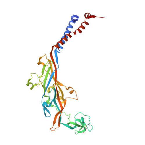
    P2X3 receptor channels expressed in sensory neurons are activated by extracellular ATP and serve important roles in nociception and sensory hypersensitization, making them attractive therapeutic targets. Although several P2X3 structures are known, it is unclear how physiologically abundant Ca 2+ -ATP and Mg 2+ -ATP activate the receptor, or how divalent cations regulate channel function. We used structural, computational and functional approaches to show that a crucial acidic chamber near the nucleotide-binding pocket in human P2X3 receptors accommodates divalent ions in two distinct modes in the absence and presence of nucleotide. The unusual engagement between the receptor, divalent ion and the γ-phosphate of ATP enables channel activation by ATP-divalent complex, cooperatively stabilizes the nucleotide on the receptor to slow ATP unbinding and recovery from desensitization, a key mechanism for limiting channel activity. These findings reveal how P2X3 receptors recognize and are activated by divalent-bound ATP, aiding future physiological investigations and drug development.


  • Organizational Affiliation

    Molecular Physiology and Biophysics Section, Porter Neuroscience Research Center, National Institute of Neurological Disorders and Stroke, National Institutes of Health, Bethesda, United States.


Macromolecules
Find similar proteins by:  (by identity cutoff)  |  3D Structure
Entity ID: 1
MoleculeChains Sequence LengthOrganismDetailsImage
P2X purinoceptor 3
A, B, C
362Homo sapiensMutation(s): 0 
Gene Names: P2RX3
Membrane Entity: Yes 
UniProt & NIH Common Fund Data Resources
Find proteins for P56373 (Homo sapiens)
Explore P56373 
Go to UniProtKB:  P56373
PHAROS:  P56373
GTEx:  ENSG00000109991 
Entity Groups  
Sequence Clusters30% Identity50% Identity70% Identity90% Identity95% Identity100% Identity
UniProt GroupP56373
Sequence Annotations
Expand
  • Reference Sequence
Oligosaccharides

Help

Entity ID: 2
MoleculeChains Length2D Diagram Glycosylation3D Interactions
alpha-D-glucopyranose-(1-4)-alpha-D-glucopyranose
D, E, F
2N/A
Glycosylation Resources
GlyTouCan:  G07411ON
GlyCosmos:  G07411ON
Small Molecules
Ligands 4 Unique
IDChains Name / Formula / InChI Key2D Diagram3D Interactions
ATP
Query on ATP

Download Ideal Coordinates CCD File 
G [auth A],
K [auth A],
Q [auth B]
ADENOSINE-5'-TRIPHOSPHATE
C10 H16 N5 O13 P3
ZKHQWZAMYRWXGA-KQYNXXCUSA-N
NAG
Query on NAG

Download Ideal Coordinates CCD File 
H [auth A]
I [auth A]
M [auth B]
N [auth B]
O [auth B]
H [auth A],
I [auth A],
M [auth B],
N [auth B],
O [auth B],
S [auth C],
T [auth C],
U [auth C]
2-acetamido-2-deoxy-beta-D-glucopyranose
C8 H15 N O6
OVRNDRQMDRJTHS-FMDGEEDCSA-N
EDO
Query on EDO

Download Ideal Coordinates CCD File 
J [auth A],
P [auth B],
V [auth C]
1,2-ETHANEDIOL
C2 H6 O2
LYCAIKOWRPUZTN-UHFFFAOYSA-N
MG
Query on MG

Download Ideal Coordinates CCD File 
L [auth A],
R [auth B],
W [auth C]
MAGNESIUM ION
Mg
JLVVSXFLKOJNIY-UHFFFAOYSA-N
Binding Affinity Annotations 
IDSourceBinding Affinity
ATP BindingDB:  6AH5 EC50: 340 (nM) from 1 assay(s)
Biologically Interesting Molecules (External Reference) 1 Unique
Experimental Data & Validation

Experimental Data

  • Method: X-RAY DIFFRACTION
  • Resolution: 3.82 Å
  • R-Value Free: 0.307 
  • R-Value Work: 0.256 
  • R-Value Observed: 0.260 
  • Space Group: C 2 2 21
Unit Cell:
Length ( Å )Angle ( ˚ )
a = 110.531α = 90
b = 139.436β = 90
c = 323.963γ = 90
Software Package:
Software NamePurpose
PHENIXrefinement
XDSdata reduction
XDSdata scaling
PHASERphasing

Structure Validation

View Full Validation Report


Ligand Structure Quality Assessment 


Entry History 

Deposition Data

  • Released Date: 2019-06-12 
  • Deposition Author(s): Hattori, M.

Revision History  (Full details and data files)

  • Version 1.0: 2019-06-12
    Type: Initial release
  • Version 2.0: 2020-07-29
    Type: Remediation
    Reason: Carbohydrate remediation
    Changes: Advisory, Atomic model, Data collection, Derived calculations, Non-polymer description, Structure summary
  • Version 2.1: 2020-12-23
    Changes: Database references, Structure summary