6AK3 | pdb_00006ak3

Crystal structure of the human prostaglandin E receptor EP3 bound to prostaglandin E2


Experimental Data Snapshot

  • Method: X-RAY DIFFRACTION
  • Resolution: 2.90 Å
  • R-Value Free: 
    0.295 (Depositor), 0.302 (DCC) 
  • R-Value Work: 
    0.251 (Depositor), 0.257 (DCC) 
  • R-Value Observed: 
    0.253 (Depositor) 

wwPDB Validation 3D Report Full Report

Validation slider image for 6AK3

Ligand Structure Quality Assessment 


This is version 1.5 of the entry. See complete history

Literature

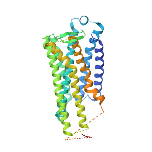
Crystal structure of the endogenous agonist-bound prostanoid receptor EP3.

Morimoto, K.Suno, R.Hotta, Y.Yamashita, K.Hirata, K.Yamamoto, M.Narumiya, S.Iwata, S.Kobayashi, T.

(2019) Nat Chem Biol 15: 8-10

  • DOI: https://doi.org/10.1038/s41589-018-0171-8
  • Primary Citation Related Structures: 
    6AK3

  • PubMed Abstract: 

    Prostanoids are a series of bioactive lipid metabolites that function in an autacoid manner via activation of cognate G-protein-coupled receptors (GPCRs). Here, we report the crystal structure of human prostaglandin (PG) E receptor subtype EP3 bound to endogenous ligand PGE 2 at 2.90 Å resolution. The structure reveals important insights into the activation mechanism of prostanoid receptors and provides a molecular basis for the binding modes of endogenous ligands.


  • Organizational Affiliation
    • Department of Cell Biology, Graduate School of Medicine, Kyoto University, Konoe-cho, Yoshida, Sakyo-ku, Kyoto, Japan. k.morimoto@mfour.med.kyoto-u.ac.jp.

Macromolecule Content 

  • Total Structure Weight: 94.31 kDa 
  • Atom Count: 4,251 
  • Modeled Residue Count: 544 
  • Deposited Residue Count: 836 
  • Unique protein chains: 1

Macromolecules

Find similar proteins by:|  3D Structure
Entity ID: 1
MoleculeChains  Sequence LengthOrganismDetailsImage
Prostaglandin E2 receptor EP3 subtype,Soluble cytochrome b562A [auth B],
B [auth A]
418Homo sapiensEscherichia coliMutation(s): 9 
Gene Names: PTGER3cybC
Membrane Entity: Yes 
UniProt & NIH Common Fund Data Resources
Find proteins for P0ABE7 (Escherichia coli)
Explore P0ABE7 
Go to UniProtKB:  P0ABE7
Find proteins for P43115 (Homo sapiens)
Explore P43115 
Go to UniProtKB:  P43115
PHAROS:  P43115
GTEx:  ENSG00000050628 
Entity Groups
Sequence Clusters30% Identity50% Identity70% Identity90% Identity95% Identity100% Identity
UniProt GroupsP0ABE7P43115
Sequence Annotations
Expand
Reference Sequence

Small Molecules

Experimental Data & Validation

Experimental Data

  • Method: X-RAY DIFFRACTION
  • Resolution: 2.90 Å
  • R-Value Free:  0.295 (Depositor), 0.302 (DCC) 
  • R-Value Work:  0.251 (Depositor), 0.257 (DCC) 
  • R-Value Observed: 0.253 (Depositor) 
Space Group: P 1 21 1
Unit Cell:
Length ( Å )Angle ( ˚ )
a = 66.09α = 90
b = 42.28β = 96.09
c = 161.3γ = 90
Software Package:
Software NamePurpose
PHENIXrefinement
XDSdata reduction
XDSdata scaling
PHASERphasing

Structure Validation

View Full Validation Report


Ligand Structure Quality Assessment 


Entry History 

& Funding Information

Deposition Data


Funding OrganizationLocationGrant Number
Japan Agency for Medical Research and Development (AMED)JapanJP18gm0910007
Japan Agency for Medical Research and Development (AMED)JapanJP18am0101079

Revision History  (Full details and data files)

  • Version 1.0: 2018-12-05
    Type: Initial release
  • Version 1.1: 2018-12-12
    Changes: Data collection, Structure summary
  • Version 1.2: 2018-12-19
    Changes: Data collection, Database references
  • Version 1.3: 2019-01-30
    Changes: Data collection, Database references
  • Version 1.4: 2023-09-06
    Changes: Data collection, Database references
  • Version 1.5: 2024-11-06
    Changes: Structure summary