6AMB | pdb_00006amb

Crystal Structure of the Afadin RA1 domain in complex with HRAS


Experimental Data Snapshot

  • Method: X-RAY DIFFRACTION
  • Resolution: 2.50 Å
  • R-Value Free: 
    0.266 (Depositor), 0.260 (DCC) 
  • R-Value Work: 
    0.218 (Depositor), 0.218 (DCC) 
  • R-Value Observed: 
    0.223 (Depositor) 

Starting Model: experimental
View more details

wwPDB Validation 3D Report Full Report

Validation slider image for 6AMB

Ligand Structure Quality Assessment 


This is version 1.2 of the entry. See complete history

Literature

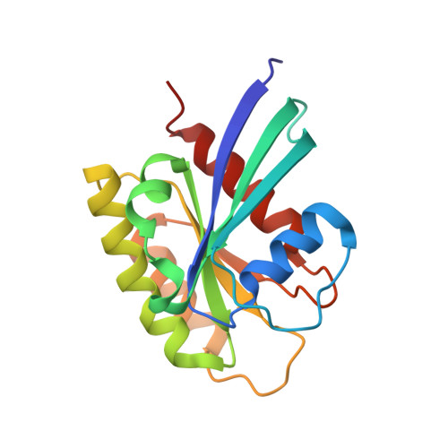
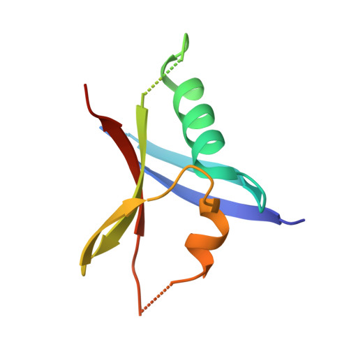
Evolution of AF6-RAS association and its implications in mixed-lineage leukemia.

Smith, M.J.Ottoni, E.Ishiyama, N.Goudreault, M.Haman, A.Meyer, C.Tucholska, M.Gasmi-Seabrook, G.Menezes, S.Laister, R.C.Minden, M.D.Marschalek, R.Gingras, A.C.Hoang, T.Ikura, M.

(2017) Nat Commun 8: 1099-1099

  • DOI: https://doi.org/10.1038/s41467-017-01326-5
  • Primary Citation Related Structures: 
    6AMB

  • PubMed Abstract: 

    Elucidation of activation mechanisms governing protein fusions is essential for therapeutic development. MLL undergoes rearrangement with numerous partners, including a recurrent translocation fusing the epigenetic regulator to a cytoplasmic RAS effector, AF6/afadin. We show here that AF6 employs a non-canonical, evolutionarily conserved α-helix to bind RAS, unique to AF6 and the classical RASSF effectors. Further, all patients with MLL-AF6 translocations express fusion proteins missing only this helix from AF6, resulting in exposure of hydrophobic residues that induce dimerization. We provide evidence that oligomerization is the dominant mechanism driving oncogenesis from rare MLL translocation partners and employ our mechanistic understanding of MLL-AF6 to examine how dimers induce leukemia. Proteomic data resolve association of dimerized MLL with gene expression modulators, and inhibiting dimerization disrupts formation of these complexes while completely abrogating leukemogenesis in mice. Oncogenic gene translocations are thus selected under pressure from protein structure/function, underscoring the complex nature of chromosomal rearrangements.


  • Organizational Affiliation
    • Institute for Research in Immunology and Cancer, Université de Montréal, Montréal, QC, H3T 1J4, Canada. matthew.james.smith@umontreal.ca.

Macromolecule Content 

  • Total Structure Weight: 31.46 kDa 
  • Atom Count: 2,016 
  • Modeled Residue Count: 249 
  • Deposited Residue Count: 269 
  • Unique protein chains: 2

Macromolecules

Find similar proteins by:|  3D Structure
Entity ID: 1
MoleculeChains  Sequence LengthOrganismDetailsImage
GTPase HRas170Homo sapiensMutation(s): 0 
Gene Names: HRASHRAS1
EC: 3.6.5.2
UniProt & NIH Common Fund Data Resources
Find proteins for P01112 (Homo sapiens)
Explore P01112 
Go to UniProtKB:  P01112
PHAROS:  P01112
GTEx:  ENSG00000174775 
Entity Groups
Sequence Clusters30% Identity50% Identity70% Identity90% Identity95% Identity100% Identity
UniProt GroupP01112
Sequence Annotations
Expand
Reference Sequence
Find similar proteins by:|  3D Structure
Entity ID: 2
MoleculeChains  Sequence LengthOrganismDetailsImage
Afadin99Mus musculusMutation(s): 0 
Gene Names: AfdnAf6Mllt4
UniProt & NIH Common Fund Data Resources
Find proteins for Q9QZQ1 (Mus musculus)
Explore Q9QZQ1 
Go to UniProtKB:  Q9QZQ1
IMPC:  MGI:1314653
Entity Groups
Sequence Clusters30% Identity50% Identity70% Identity90% Identity95% Identity100% Identity
UniProt GroupQ9QZQ1
Sequence Annotations
Expand
Reference Sequence

Small Molecules

Ligands 2 Unique
IDChains Name / Formula / InChI Key2D Diagram3D Interactions
GNP

Query on GNP


Download:Ideal Coordinates CCD File
C [auth A]PHOSPHOAMINOPHOSPHONIC ACID-GUANYLATE ESTER
C10 H17 N6 O13 P3
UQABYHGXWYXDTK-UUOKFMHZSA-N
MG

Query on MG


Download:Ideal Coordinates CCD File
D [auth A]MAGNESIUM ION
Mg
JLVVSXFLKOJNIY-UHFFFAOYSA-N

Experimental Data & Validation

Experimental Data

  • Method: X-RAY DIFFRACTION
  • Resolution: 2.50 Å
  • R-Value Free:  0.266 (Depositor), 0.260 (DCC) 
  • R-Value Work:  0.218 (Depositor), 0.218 (DCC) 
  • R-Value Observed: 0.223 (Depositor) 
Space Group: P 21 21 21
Unit Cell:
Length ( Å )Angle ( ˚ )
a = 48.239α = 90
b = 57.152β = 90
c = 73.329γ = 90
Software Package:
Software NamePurpose
HKL-2000data scaling
PHENIXrefinement
PDB_EXTRACTdata extraction
HKL-2000data reduction
PHASERphasing

Structure Validation

View Full Validation Report


Ligand Structure Quality Assessment 


Entry History 

Deposition Data

Revision History  (Full details and data files)

  • Version 1.0: 2017-11-01
    Type: Initial release
  • Version 1.1: 2017-11-08
    Changes: Database references
  • Version 1.2: 2023-10-04
    Changes: Data collection, Database references, Refinement description