6B98 | pdb_00006b98

PDE2 in complex with compound 1


Experimental Data Snapshot

  • Method: X-RAY DIFFRACTION
  • Resolution: 1.97 Å
  • R-Value Free: 
    0.240 (Depositor), 0.248 (DCC) 
  • R-Value Work: 
    0.199 (Depositor) 
  • R-Value Observed: 
    0.202 (Depositor) 

wwPDB Validation 3D Report Full Report

Validation slider image for 6B98

Ligand Structure Quality Assessment 


This is version 1.2 of the entry. See complete history

Literature

The identification of a novel lead class for phosphodiesterase 2 inhibition by fragment-based drug design.

Forster, A.B.Abeywickrema, P.Bunda, J.Cox, C.D.Cabalu, T.D.Egbertson, M.Fay, J.Getty, K.Hall, D.Kornienko, M.Lu, J.Parthasarathy, G.Reid, J.Sharma, S.Shipe, W.D.Smith, S.M.Soisson, S.Stachel, S.J.Su, H.P.Wang, D.Berger, R.

(2017) Bioorg Med Chem Lett 27: 5167-5171

  • DOI: https://doi.org/10.1016/j.bmcl.2017.10.054
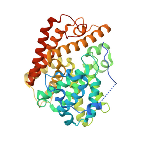
  • Primary Citation Related Structures: 
    6B96, 6B97, 6B98

  • PubMed Abstract: 

    We have identified a novel PDE2 inhibitor series using fragment-based screening. Pyrazolopyrimidine fragment 1, while possessing weak potency (K i  = 22.4 μM), exhibited good binding efficiencies (LBE = 0.49, LLE = 4.48) to serve as a start for structure-based drug design. With the assistance of molecular modeling and X-ray crystallography, this fragment was developed into a series of potent PDE2 inhibitors with good physicochemical properties. Compound 16, a PDE2 selective inhibitor, was identified that exhibited favorable rat pharmacokinetic properties.


  • Organizational Affiliation
    • Discovery Chemistry, MRL, 770 Sumneytown Pike, West Point, PA 19486, USA. Electronic address: Ashley_nomland@merck.com.

Macromolecule Content 

  • Total Structure Weight: 87.96 kDa 
  • Atom Count: 5,833 
  • Modeled Residue Count: 693 
  • Deposited Residue Count: 746 
  • Unique protein chains: 1

Macromolecules

Find similar proteins by:|  3D Structure
Entity ID: 1
MoleculeChains  Sequence LengthOrganismDetailsImage
cGMP-dependent 3',5'-cyclic phosphodiesterase
A, B
373Homo sapiensMutation(s): 0 
Gene Names: PDE2A
EC: 3.1.4.17
UniProt & NIH Common Fund Data Resources
Find proteins for O00408 (Homo sapiens)
Explore O00408 
Go to UniProtKB:  O00408
PHAROS:  O00408
GTEx:  ENSG00000186642 
Entity Groups
Sequence Clusters30% Identity50% Identity70% Identity90% Identity95% Identity100% Identity
UniProt GroupO00408
Sequence Annotations
Expand
Reference Sequence

Small Molecules

Binding Affinity Annotations 
IDSourceBinding Affinity
D07 BindingDB:  6B98 Ki: 2.24e+4 (nM) from 1 assay(s)

Experimental Data & Validation

Experimental Data

  • Method: X-RAY DIFFRACTION
  • Resolution: 1.97 Å
  • R-Value Free:  0.240 (Depositor), 0.248 (DCC) 
  • R-Value Work:  0.199 (Depositor) 
  • R-Value Observed: 0.202 (Depositor) 
Space Group: P 21 21 21
Unit Cell:
Length ( Å )Angle ( ˚ )
a = 72.034α = 90
b = 94.186β = 90
c = 102.526γ = 90
Software Package:
Software NamePurpose
BUSTERrefinement
XDSdata reduction
autoPROCdata scaling

Structure Validation

View Full Validation Report


Ligand Structure Quality Assessment 


Entry History 

Deposition Data

  • Released Date: 2017-11-22 
  • Deposition Author(s): Lu, J.

Revision History  (Full details and data files)

  • Version 1.0: 2017-11-22
    Type: Initial release
  • Version 1.1: 2017-11-29
    Changes: Database references
  • Version 1.2: 2024-03-06
    Changes: Data collection, Database references, Derived calculations, Refinement description