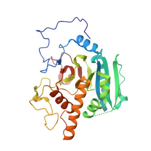
Conserved residues Arg188 and Asp302 are critical for active site organization and catalysis in human ABO(H) blood group A and B glycosyltransferases.
Gagnon, S.M.L., Legg, M.S.G., Polakowski, R., Letts, J.A., Persson, M., Lin, S., Zheng, R.B., Rempel, B., Schuman, B., Haji-Ghassemi, O., Borisova, S.N., Palcic, M.M., Evans, S.V.(2018) Glycobiology 28: 624-636
- PubMed: 29873711
- DOI: https://doi.org/10.1093/glycob/cwy051
- Primary Citation of Related Structures:
6BJI, 6BJJ, 6BJK, 6BJL, 6BJM - PubMed Abstract:
Homologous glycosyltransferases GTA and GTB perform the final step in human ABO(H) blood group A and B antigen synthesis by transferring the sugar moiety from donor UDP-GalNAc/UDP-Gal to the terminal H antigen disaccharide acceptor. Like other GT-A fold family 6 glycosyltransferases, GTA and GTB undergo major conformational changes in two mobile regions, the C-terminal tail and internal loop, to achieve the closed, catalytic state. These changes are known to establish a salt bridge network among conserved active site residues Arg188, Asp211 and Asp302, which move to accommodate a series of discrete donor conformations while promoting loop ordering and formation of the closed enzyme state. However, the individual significance of these residues in linking these processes remains unclear. Here, we report the kinetics and high-resolution structures of GTA/GTB mutants of residues 188 and 302. The structural data support a conserved salt bridge network critical to mobile polypeptide loop organization and stabilization of the catalytically competent donor conformation. Consistent with the X-ray crystal structures, the kinetic data suggest that disruption of this salt bridge network has a destabilizing effect on the transition state, emphasizing the importance of Arg188 and Asp302 in the glycosyltransfer reaction mechanism. The salt bridge network observed in GTA/GTB structures during substrate binding appears to be conserved not only among other Carbohydrate Active EnZyme family 6 glycosyltransferases but also within both retaining and inverting GT-A fold glycosyltransferases. Our findings augment recently published crystal structures, which have identified a correlation between donor substrate conformational changes and mobile loop ordering.
Organizational Affiliation:
Department of Biochemistry & Microbiology, University of Victoria, STN CSC, Victoria, BC, Canada.


