6C7F | pdb_00006c7f

Crystal structure of human phosphodiesterase 2A with 1-(2-chloro-5-isobutoxy-phenyl)-N,4-dimethyl-[1,2,4]triazolo[4,3-a]quinoxaline-8-carboxamide


Experimental Data Snapshot

  • Method: X-RAY DIFFRACTION
  • Resolution: 1.82 Å
  • R-Value Free: 
    0.246 (Depositor) 
  • R-Value Work: 
    0.195 (Depositor) 
  • R-Value Observed: 
    0.198 (Depositor) 

wwPDB Validation 3D Report Full Report

Validation slider image for 6C7F

Ligand Structure Quality Assessment 


This is version 1.3 of the entry. See complete history

Literature

Mathematical and Structural Characterization of Strong Nonadditive Structure-Activity Relationship Caused by Protein Conformational Changes.

Gomez, L.Xu, R.Sinko, W.Selfridge, B.Vernier, W.Ly, K.Truong, R.Metz, M.Marrone, T.Sebring, K.Yan, Y.Appleton, B.Aertgeerts, K.Massari, M.E.Breitenbucher, J.G.

(2018) J Med Chem 61: 7754-7766

  • DOI: https://doi.org/10.1021/acs.jmedchem.8b00713
  • Primary Citation Related Structures: 
    6C7D, 6C7E, 6C7F, 6C7G, 6C7I, 6C7J

  • PubMed Abstract: 

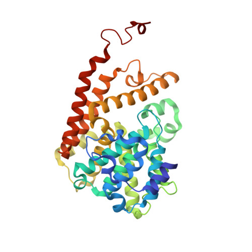
    In medicinal chemistry, accurate prediction of additivity-based structure-activity relationship (SAR) analysis rests on three assumptions: (1) a consistent binding pose of the central scaffold, (2) no interaction between the R group substituents, and (3) a relatively rigid binding pocket in which the R group substituents act independently. Previously, examples of nonadditive SAR have been documented in systems that deviate from the first two assumptions. Local protein structural change upon ligand binding, through induced fit or conformational selection, although a well-known phenomenon that invalidates the third assumption, has not been linked to nonadditive SAR conclusively. Here, for the first time, we present clear structural evidence that the formation of a hydrophobic pocket upon ligand binding in PDE2 catalytic site reduces the size of another distinct subpocket and contributes to strong nonadditive SAR between two otherwise distant R groups.


  • Organizational Affiliation
    • Dart Neuroscience LLC , 12278 Scripps Summit Drive , San Diego , California 92131 , United States.

Macromolecule Content 

  • Total Structure Weight: 121.27 kDa 
  • Atom Count: 8,814 
  • Modeled Residue Count: 994 
  • Deposited Residue Count: 1,026 
  • Unique protein chains: 1

Macromolecules

Find similar proteins by:|  3D Structure
Entity ID: 1
MoleculeChains  Sequence LengthOrganismDetailsImage
cGMP-dependent 3',5'-cyclic phosphodiesterase
A, B, C
342Homo sapiensMutation(s): 0 
Gene Names: PDE2A
EC: 3.1.4.17
UniProt & NIH Common Fund Data Resources
Find proteins for O00408 (Homo sapiens)
Explore O00408 
Go to UniProtKB:  O00408
PHAROS:  O00408
GTEx:  ENSG00000186642 
Entity Groups
Sequence Clusters30% Identity50% Identity70% Identity90% Identity95% Identity100% Identity
UniProt GroupO00408
Sequence Annotations
Expand
Reference Sequence

Small Molecules

Ligands 3 Unique
IDChains Name / Formula / InChI Key2D Diagram3D Interactions
EOS

Query on EOS


Download:Ideal Coordinates CCD File
D [auth A],
G [auth B],
J [auth C]
1-[2-chloro-5-(2-methylpropoxy)phenyl]-N,4-dimethyl[1,2,4]triazolo[4,3-a]quinoxaline-8-carboxamide
C22 H22 Cl N5 O2
NUXSVUZDEZKZLN-UHFFFAOYSA-N
ZN

Query on ZN


Download:Ideal Coordinates CCD File
E [auth A],
H [auth B],
K [auth C]
ZINC ION
Zn
PTFCDOFLOPIGGS-UHFFFAOYSA-N
MG

Query on MG


Download:Ideal Coordinates CCD File
F [auth A],
I [auth B],
L [auth C]
MAGNESIUM ION
Mg
JLVVSXFLKOJNIY-UHFFFAOYSA-N

Experimental Data & Validation

Experimental Data

  • Method: X-RAY DIFFRACTION
  • Resolution: 1.82 Å
  • R-Value Free:  0.246 (Depositor) 
  • R-Value Work:  0.195 (Depositor) 
  • R-Value Observed: 0.198 (Depositor) 
Space Group: C 1 2 1
Unit Cell:
Length ( Å )Angle ( ˚ )
a = 167.248α = 90
b = 73.004β = 110.33
c = 91.953γ = 90
Software Package:
Software NamePurpose
DIALSdata reduction
Aimlessdata scaling
PHASERphasing
PHENIXrefinement
PDB_EXTRACTdata extraction

Structure Validation

View Full Validation Report


Ligand Structure Quality Assessment 


Entry History 

Deposition Data

Revision History  (Full details and data files)

  • Version 1.0: 2018-08-15
    Type: Initial release
  • Version 1.1: 2018-08-29
    Changes: Data collection, Database references
  • Version 1.2: 2018-09-26
    Changes: Data collection, Database references
  • Version 1.3: 2024-03-13
    Changes: Data collection, Database references, Derived calculations